Volume 29, Number 7—July 2023
Dispatch
Cutaneous Pythiosis in 2 Dogs, Italy
Abstract
We report cutaneous pythiosis in 2 dogs in Italy that had recurrent exposure to the same freshwater habitat. Phylogenetic analysis placed the isolates within Pythium insidiosum complex cluster IV, corresponding to P. periculosum. In Italy, pythiosis should be considered in differential diagnoses by human and veterinary health professionals.
Pythiosis is a granulomatous disease caused by oomycete organisms affecting mainly horses, humans, and dogs, and is historically associated with humid environments (1). In India, where the first cases were reported in horses in the late 19th century, pythiosis is known as bursattee, from burus, signifying rain, because infections appeared during the rainy season (2). In other countries, pythiosis is known by different names, such as swamp cancer in Australia and the United States and horse leeches in the United States (2).
The genus Pythium (kingdom Stramenopila) comprises >120 species inhabiting all soil and wet environments. Pythium spp. have a saprophytic life cycle, although many are plant pathogens (3). P. insidiosum has been considered the only species that infects mammals (2). In 2003, existence of a cryptic species was suspected on the basis of phylogenetic analyses that indicated P. insidiosum is a complex with 3 clusters, 1 (cluster III) of which displays substantial divergence from the other 2 clusters (4). Recent studies have revealed a fourth cluster (cluster IV); clusters III and IV form a monophyletic group representing a novel species, P. periculosum (5). P. aphanidermatum has also been reported in 2 cases of human infection (6,7).
Pythiosis is characterized by the presence of broad, irregular, perpendicular branching hyphae in tissues and cultures that are aseptate in early growth stages or sparsely septate in aged organisms. Biflagellate motile zoospores are the infecting agents, and production in the environment requires free water. The zoospores are attracted by open wounds, encyst on exposed tissue, and develop a germ tube that mechanically penetrates tissue (2). The optimal growth temperature range for zoospores is 28°C–37°C (3). Pythiosis manifests as rapidly growing granulomatous lesions that might be devastating and life-threatening (2). Organs and tissues most affected are the skin, gastrointestinal tract, eyes, and blood vessels (2,3); disseminated forms are also possible (3).
Pythiosis is diffused in warm and humid areas of tropical and subtropical countries, including northeastern Australia, Brazil, Colombia, Costa Rica, India, Thailand, Uruguay, southern and southeastern states of the United States, and Venezuela. The disease has also been reported sporadically in some temperate regions (3). A horse with compatible clinical pythiosis features was described in France in 1896 (8), whereas confirmed cases have been recently reported in human patients in Spain (9,10). We describe 2 cases of cutaneous pythiosis in dogs in Italy.
Figure 1
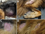
Figure 1. Clinical manifestation of cutaneous pythiosis in 2 dogs, Italy. A, B) Case 1, showing a large mass on the right flank (A) and a large mass on the left axilla...
We evaluated soft tissue swellings in 2 unrelated dogs that lived 30 km apart in Rome province of central Italy in October 2022 (case 1) and January 2023 (case 2) (Figure 1). The animals had never been outside of Italy before onset of skin lesions and were otherwise healthy. We collected anamnesis and clinical manifestation data (Table).
Figure 2
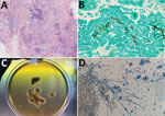
Figure 2. Histological analyses of cutaneous pythiosis in 2 dogs, Italy. A) Section from punch biopsy of cutaneous mass showing confluent pyogranulomatous and eosinophilic granulomas with central necrosis in the deep dermis...
Histopathologic results of lesion punch biopsies showed multifocal to coalescing (pyo)granulomatous and eosinophilic dermatitis and panniculitis with intralesional, irregular branching hyphae (Figure 2, panel A). The hyphae were positive for Grocott methenamine silver stain, appearing dark brown (Figure 2, panel B), whereas they were negative after staining with periodic acid Schiff stain. Culturing on Sabouraud dextrose agar (for fungi) containing chloramphenicol and gentamycin yielded negative results.
To obtain a definitive identification, we extracted genomic DNA from biopsied tissues by using a NucleoSpin Tissue kit (Macherey-Nagel, https://www.mn-net.com). We PCR amplified and sequenced the internal transcribed spacer (ITS) region by using the primer pair ITS4 and ITS5 (11). We obtained identical sequences (GenBank accession nos. OQ532907 [case 1] and OQ532908 [case 2]) that were closely related to ITS sequences of P. insidiosum from GenBank (compared by using BLASTn, https://blast.ncbi.nlm.nih.gov). We aligned sequences belonging to different clusters within the P. insidiosum complex (5) by using MEGA11 software (https://www.megasoftware.net). We conducted phylogenetic analysis by using the neighbor-joining method (bootstrap analysis with 1,000 replicates); sequences obtained from the infected dog tissues clustered with Pythium cluster IV (Appendix Figure), corresponding to the newly described species P. periculosum (5).
Although an environmental study would be necessary for confirmation, Lake Bracciano near Rome might have been the infection source (Table). Both dogs regularly swam in the lake, which has vast stagnant waters with stable temperatures of ≈30°C in hotter months, features known to support pathogenic Pythium spp. growth (2,3).
Additional material was collected from the lesion in case 2 through fine-needle aspiration. We suspected the organism failed to grow in previous cultures because of chloramphenicol supplementation (10). Therefore, we cultured the aspirate on unsupplemented Sabouraud dextrose agar at 37°C. After a 24-hour incubation, submerged, colorless colonies with irregular radiate patterns developed from the aspirated material (Figure 2, panel C). Microscopically, the hyphae were broad (4–10 µm in diameter), hyaline, and sparsely septate (Figure 2, panel D). Colonies were identified as P. periculosum by using the same molecular approach applied to biopsies.
Our cases suggest a geographic distribution of pythiosis broader than previously recognized, aligning with other reports of the disease outside of classical tropical and subtropical areas, such as US regions near the border of Canada, as well as Spain, Israel, Japan, and South Korea (2,3). Because the average climatic conditions in temperate zones appear unsuitable for the causative oomycete life cycle, cases in those areas likely occur in restricted ecologic niches. Our report illustrates that concept; central Italy has a primarily Mediterranean climate that has mild, sometimes rainy winters and sunny, hot, and usually dry summers. Those features do not fit the description of areas more prone to pythiosis. However, the zone where we suspect the dogs became infected has features of a pythiosis-risk area (warm, humid environment). Increasing reports of pythiosis in nonendemic regions might indicate an expansion of the causative oomycetes because of global climate changes. Another interpretation is that more cases are recognized and published because of increased awareness of healthcare personnel and availability of diagnostic tools (3). Environmental and clinical studies will be necessary to address those hypotheses.
The dogs in this report showed signs typically associated with the cutaneous form of pythiosis, including developing multiple masses or dermal plaques over time (3). Gastrointestinal pythiosis in dogs is another form of the disease characterized by weight loss, vomiting, diarrhea, and hematochezia (3,12).
Genetic variation associated with geographic provenance exists for species within the P. insidiosum complex (4,5). P. insidiosum cluster I is mainly found throughout the Americas, whereas P. insidiosum cluster II is typically found in Asia and Australia. P. periculosum cluster III has been reported mainly in the United States and is sympatric with members of cluster I (5). The genotype identified in our cases (P. periculosum cluster IV) seems to have a broader distribution; most reports of human cases have been from Thailand and India (3). However, P. periculosum cluster IV has also been recovered from environmental samples in Brazil and the United States (13,14) and from human patients in Israel (15) and Spain (10), which is noteworthy because of the close proximity of Spain to Italy.
In conclusion, from a One Health perspective, our study shows the environmental presence of an unexpected, exotic pathogen that could potentially infect humans in a country with a temperate climate. In Italy, pythiosis should now be considered a differential diagnosis by human and veterinary health professionals, especially in cases where there is a history of exposure to freshwater habitats.
Dr. Peano is an assistant professor at the Department of Veterinary Sciences, Parasitology Section, University of Turin, in northwest Italy. His primary research interests are parasitic and fungal diseases of domestic and wild animals.
Acknowledgment
We thank the dogs’ owners for consenting to the publication of the cases and Gaetano Reggi for referring 1 case.
References
- De Cock AW, Mendoza L, Padhye AA, Ajello L, Kaufman L. Pythium insidiosum sp. nov., the etiologic agent of pythiosis. J Clin Microbiol. 1987;25:344–9. DOIPubMedGoogle Scholar
- Gaastra W, Lipman LJA, De Cock AW, Exel TK, Pegge RBG, Scheurwater J, et al. Pythium insidiosum: an overview. Vet Microbiol. 2010;146:1–16. DOIPubMedGoogle Scholar
- Yolanda H, Krajaejun T. Global distribution and clinical features of pythiosis in humans and animals. J Fungi (Basel). 2022;8:182. DOIPubMedGoogle Scholar
- Schurko AM, Mendoza L, Lévesque CA, Désaulniers NL, de Cock AW, Klassen GR. A molecular phylogeny of Pythium insidiosum. Mycol Res. 2003;107:537–44. DOIPubMedGoogle Scholar
- Miraglia BM, Mendoza L, Rammohan R, Vilela L, Vilela C, Vilela G, et al. Pythium insidiosum complex hides a cryptic novel species: Pythium periculosum. Fungal Biol. 2022;126:366–74. DOIPubMedGoogle Scholar
- Calvano TP, Blatz PJ, Vento TJ, Wickes BL, Sutton DA, Thompson EH, et al. Pythium aphanidermatum infection following combat trauma. J Clin Microbiol. 2011;49:3710–3. DOIPubMedGoogle Scholar
- Farmer AR, Murray CK, Driscoll IR, Wickes BL, Wiederhold N, Sutton DA, et al. Combat-related Pythium aphanidermatum invasive wound infection: case report and discussion of utility of molecular diagnostics. J Clin Microbiol. 2015;53:1968–75. DOIPubMedGoogle Scholar
- Drouin V. Sur une nouvelle mycose du cheval. Rec Med Vet. 1896;30:337–44.
- Del Castillo-Jiménez MC, Baptista-Díaz N, Montero J, Pascual A. [Pythium insidiosum ocular infection] [in Spanish]. Enferm Infecc Microbiol Clin. 2013;31:118–9. DOIPubMedGoogle Scholar
- Bernheim D, Dupont D, Aptel F, Dard C, Chiquet C, Normand AC, et al. Pythiosis: Case report leading to new features in clinical and diagnostic management of this fungal-like infection. Int J Infect Dis. 2019;86:40–3. DOIPubMedGoogle Scholar
- White TJ, Bruns T, Lee S, Taylor J. Amplification and direct sequencing of fungal ribosomal RNA genes for phylogenetics. In: Innis MA, Gelfand DH, Sninsky JJ, White TJ, editors. PCR protocols: a guide to methods and applications. San Diego (CA): Academic Press; 1990. p. 315–22.
- Berryessa NA, Marks SL, Pesavento PA, Krasnansky T, Yoshimoto SK, Johnson EG, et al. Gastrointestinal pythiosis in 10 dogs from California. J Vet Intern Med. 2008;22:1065–9. DOIPubMedGoogle Scholar
- Paz GSD, Camargo GG, Cury JE, Apolonio EVP, Garces HG, Prado ACD, et al. Outbreak of equine pythiosis in a southeastern region of Brazil: Environmental isolation and phylogeny. Transbound Emerg Dis. 2022;69:1617–24. DOIPubMedGoogle Scholar
- Presser JW, Goss EM. Environmental sampling reveals that Pythium insidiosum is ubiquitous and genetically diverse in North Central Florida. Med Mycol. 2015;53:674–83. DOIPubMedGoogle Scholar
- Tanhehco TY, Stacy RC, Mendoza L, Durand ML, Jakobiec FA, Colby KA. Pythium insidiosum keratitis in Israel. Eye Contact Lens. 2011;37:96–8. DOIPubMedGoogle Scholar
Figures
Table
Cite This ArticleOriginal Publication Date: June 12, 2023
Table of Contents – Volume 29, Number 7—July 2023
| EID Search Options |
|---|
|
|
|
|
|
|
Please use the form below to submit correspondence to the authors or contact them at the following address:
Andrea Peano, Dipartimento di Scienze Veterinarie, Università di Torino, Largo P. Braccini 2, 10095 Grugliasco, Torino, Italy
Top