Synopses
Multicentric Case Series and Literature Review of Coccidioidal Otomastoiditis [PDF - 966 KB - 5 pages]
Coccidioidomycosis involving the ear, mastoid bone, or both is uncommon. We describe 5 new cases from the United States and review 4 cases reported in the literature of otomycosis and mastoiditis caused by Coccidioides. Of the 9 cases, 8 were linked to residence in or travel to California. Two patients had poorly controlled diabetes mellitus, 7 had otomastoiditis, 1 had otitis externa without mastoid involvement, and 1 had mastoiditis without otic involvement. Four patients had concurrent or prior pulmonary coccidioidomycosis. Ipsilateral facial nerve palsies developed in 2 patients. All patients received antifungal treatment for varying durations, and 8 of the 9 patients underwent surgical debridement. Clinicians should consider coccidioidomycosis as a differential diagnosis for otomastoiditis in patients with geographic risks.
| EID | Schwartz IS, Marek C, Sandhu H, Abdelmonem A, Petersen G, Dishner E, et al. Multicentric Case Series and Literature Review of Coccidioidal Otomastoiditis. Emerg Infect Dis. 2023;29(7):1297-1301. https://doi.org/10.3201/eid2907.230129 |
|---|---|
| AMA | Schwartz IS, Marek C, Sandhu H, et al. Multicentric Case Series and Literature Review of Coccidioidal Otomastoiditis. Emerging Infectious Diseases. 2023;29(7):1297-1301. doi:10.3201/eid2907.230129. |
| APA | Schwartz, I. S., Marek, C., Sandhu, H., Abdelmonem, A., Petersen, G., Dishner, E....Thompson, G. R. (2023). Multicentric Case Series and Literature Review of Coccidioidal Otomastoiditis. Emerging Infectious Diseases, 29(7), 1297-1301. https://doi.org/10.3201/eid2907.230129. |
Nationwide Outbreak of Candida auris Infections Driven by COVID-19 Hospitalizations, Israel, 2021–2022 [PDF - 3.17 MB - 10 pages]
We report an outbreak of Candida auris across multiple healthcare facilities in Israel. For the period of May 2014–May 2022, a total of 209 patients with C. auris infection or colonization were identified. The C. auris incidence rate increased 30-fold in 2021 (p = 0.00015), corresponding in time with surges of COVID-19–related hospitalization. Multilocus sequence typing revealed hospital-level outbreaks with distinct clones. A clade III clone, imported into Israel in 2016, accounted for 48.8% of typed isolates after January 2021 and was more frequently resistant to fluconazole (100% vs. 63%; p = 0.00017) and voriconazole (74% vs. 5.2%; p<0.0001) than were non–clade III isolates. A total of 23% of patients had COVID-19, and 78% received mechanical ventilation. At the hospital level, outbreaks initially involved mechanically ventilated patients in specialized COVID-19 units and then spread sequentially to ventilated non–COVID-19 patients and nonventilated patients.
| EID | Biran R, Cohen R, Finn T, Brosh-Nissimov T, Rahav G, Yahav D, et al. Nationwide Outbreak of Candida auris Infections Driven by COVID-19 Hospitalizations, Israel, 2021–2022. Emerg Infect Dis. 2023;29(7):1297-1301. https://doi.org/10.3201/eid2907.221888 |
|---|---|
| AMA | Biran R, Cohen R, Finn T, et al. Nationwide Outbreak of Candida auris Infections Driven by COVID-19 Hospitalizations, Israel, 2021–2022. Emerging Infectious Diseases. 2023;29(7):1297-1301. doi:10.3201/eid2907.221888. |
| APA | Biran, R., Cohen, R., Finn, T., Brosh-Nissimov, T., Rahav, G., Yahav, D....Ben-Ami, R. (2023). Nationwide Outbreak of Candida auris Infections Driven by COVID-19 Hospitalizations, Israel, 2021–2022. Emerging Infectious Diseases, 29(7), 1297-1301. https://doi.org/10.3201/eid2907.221888. |
Research
We retrospectively reviewed consecutive cases of mucormycosis reported from a tertiary-care center in India to determine the clinical and mycologic characteristics of emerging Rhizopus homothallicus fungus. The objectives were ascertaining the proportion of R. homothallicus infection and the 30-day mortality rate in rhino-orbital mucormycosis attributable to R. homothallicus compared with R. arrhizus. R. homothallicus accounted for 43 (6.8%) of the 631 cases of mucormycosis. R. homothallicus infection was independently associated with better survival (odds ratio [OR] 0.08 [95% CI 0.02–0.36]; p = 0.001) than for R. arrhizus infection (4/41 [9.8%] vs. 104/266 [39.1%]) after adjusting for age, intracranial involvement, and surgery. We also performed antifungal-susceptibility testing, which indicated a low range of MICs for R. homothallicus against the commonly used antifungals (amphotericin B [0.03–16], itraconazole [0.03–16], posaconazole [0.03–8], and isavuconazole [0.03–16]). 18S gene sequencing and amplified length polymorphism analysis revealed distinct clustering of R. homothallicus.
| EID | Rudramurthy SM, Singh S, Kanaujia R, Chaudhary H, Muthu V, Panda N, et al. Clinical and Mycologic Characteristics of Emerging Mucormycosis Agent Rhizopus homothallicus. Emerg Infect Dis. 2023;29(7):1313-1322. https://doi.org/10.3201/eid2907.221491 |
|---|---|
| AMA | Rudramurthy SM, Singh S, Kanaujia R, et al. Clinical and Mycologic Characteristics of Emerging Mucormycosis Agent Rhizopus homothallicus. Emerging Infectious Diseases. 2023;29(7):1313-1322. doi:10.3201/eid2907.221491. |
| APA | Rudramurthy, S. M., Singh, S., Kanaujia, R., Chaudhary, H., Muthu, V., Panda, N....Chakrabarti, A. (2023). Clinical and Mycologic Characteristics of Emerging Mucormycosis Agent Rhizopus homothallicus. Emerging Infectious Diseases, 29(7), 1313-1322. https://doi.org/10.3201/eid2907.221491. |
Trajectory and Demographic Correlates of Antibodies to SARS-CoV-2 Nucleocapsid in Recently Infected Blood Donors, United States [PDF - 2.18 MB - 7 pages]
We evaluated antibodies to the nucleocapsid protein of SARS-CoV-2 in a large cohort of blood donors in the United States who were recently infected with the virus. Antibodies to the nucleocapsid protein of SARS-CoV-2 indicate previous infection but are subject to waning, potentially affecting epidemiologic studies. We longitudinally evaluated a cohort of 19,323 blood donors who had evidence of recent infection by using a widely available serologic test to determine the dynamics of such waning. We analyzed overall signal-to-cutoff values for 48,330 donations (average 2.5 donations/person) that had an average observation period of 102 days. The observed peak signal-to-cutoff value varied widely, but the waning rate was consistent across the range, with a half-life of 122 days. Within the cohort, only 0.75% of persons became seronegative. Factors predictive of higher peak values and longer time to seroreversion included increasing age, male sex, higher body mass index, and non-Caucasian race.
| EID | Haynes JM, Dodd RY, Crowder LA, Notari EP, Stramer SL. Trajectory and Demographic Correlates of Antibodies to SARS-CoV-2 Nucleocapsid in Recently Infected Blood Donors, United States. Emerg Infect Dis. 2023;29(7):1323-1329. https://doi.org/10.3201/eid2907.230173 |
|---|---|
| AMA | Haynes JM, Dodd RY, Crowder LA, et al. Trajectory and Demographic Correlates of Antibodies to SARS-CoV-2 Nucleocapsid in Recently Infected Blood Donors, United States. Emerging Infectious Diseases. 2023;29(7):1323-1329. doi:10.3201/eid2907.230173. |
| APA | Haynes, J. M., Dodd, R. Y., Crowder, L. A., Notari, E. P., & Stramer, S. L. (2023). Trajectory and Demographic Correlates of Antibodies to SARS-CoV-2 Nucleocapsid in Recently Infected Blood Donors, United States. Emerging Infectious Diseases, 29(7), 1323-1329. https://doi.org/10.3201/eid2907.230173. |
Zoonotic outbreaks of sporotrichosis are increasing in Brazil. We examined and described the emergence of cat-transmitted sporotrichosis (CTS) caused by the fungal pathogen Sporothrix brasiliensis. We calculated incidence and mapped geographic distribution of cases in Curitiba, Brazil, by reviewing medical records from 216 sporotrichosis cases diagnosed during 2011–May 2022. Proven sporotrichosis was established in 84 (39%) patients and probable sporotrichosis in 132 (61%). Incidence increased from 0.3 cases/100,000 outpatient visit-years in 2011 to 21.4 cases/100,000 outpatient visit-years in 2021; of the 216 cases, 58% (n = 126) were diagnosed during 2019–2021. The main clinical form of sporotrichosis was lymphocutaneous (63%), followed by localized cutaneous (24%), ocular (10%), multisite infections (3%), and cutaneous disseminated (<0.5%). Since the first report of CTS in Curitiba in 2011, sporotrichosis has increased substantially, indicating continuous disease transmission. Clinician and public awareness of CTS and efforts to prevent transmission are needed.
| EID | Cognialli R, Cáceres DH, Bastos F, Cavassin FB, Lustosa B, Vicente VA, et al. Rising Incidence of Sporothrix brasiliensis Infections, Curitiba, Brazil, 2011–2022. Emerg Infect Dis. 2023;29(7):1330-1339. https://doi.org/10.3201/eid2907.230155 |
|---|---|
| AMA | Cognialli R, Cáceres DH, Bastos F, et al. Rising Incidence of Sporothrix brasiliensis Infections, Curitiba, Brazil, 2011–2022. Emerging Infectious Diseases. 2023;29(7):1330-1339. doi:10.3201/eid2907.230155. |
| APA | Cognialli, R., Cáceres, D. H., Bastos, F., Cavassin, F. B., Lustosa, B., Vicente, V. A....Queiroz-Telles, F. (2023). Rising Incidence of Sporothrix brasiliensis Infections, Curitiba, Brazil, 2011–2022. Emerging Infectious Diseases, 29(7), 1330-1339. https://doi.org/10.3201/eid2907.230155. |
Triplex ELISA for Assessing Durability of Taenia solium Seropositivity after Neurocysticercosis Cure [PDF - 2.09 MB - 9 pages]
Neurocysticercosis prevalence estimates often are based on serosurveys. However, assessments of Taenia solium seropositivity durability in patients with various neurocysticercosis types are lacking. We optimized a triplex serologic ELISA by using synthetic GP50, T24H, and Ts18var3 antigens for T. solium. We used that assay to test sequential serologic responses over several years after neurocysticercosis cure in 46 patients, 9 each with parenchymal or ventricular neurocysticercosis and 28 with subarachnoid disease. Triplex results were concordant with 98% of positive and 100% of negative enzyme-linked immunoelectrotransfer blots. Eight years after neurocysticercosis cure, 11.1% of patients with parenchymal, 47.3% with subarachnoid, and 41.7% with ventricular disease were still seropositive. Median time to seroreversion after cure in this cohort in a T. solium nonendemic area was 2 years for parenchymal disease, 4 years for ventricular disease, and 8 years for subarachnoid disease. Our findings can inform epidemiologic models that rely on serosurveys to estimate disease burden.
| EID | Tang NL, Nash TE, Corda M, Nutman TB, O’Connell EM. Triplex ELISA for Assessing Durability of Taenia solium Seropositivity after Neurocysticercosis Cure. Emerg Infect Dis. 2023;29(7):1340-1348. https://doi.org/10.3201/eid2907.230364 |
|---|---|
| AMA | Tang NL, Nash TE, Corda M, et al. Triplex ELISA for Assessing Durability of Taenia solium Seropositivity after Neurocysticercosis Cure. Emerging Infectious Diseases. 2023;29(7):1340-1348. doi:10.3201/eid2907.230364. |
| APA | Tang, N. L., Nash, T. E., Corda, M., Nutman, T. B., & O’Connell, E. M. (2023). Triplex ELISA for Assessing Durability of Taenia solium Seropositivity after Neurocysticercosis Cure. Emerging Infectious Diseases, 29(7), 1340-1348. https://doi.org/10.3201/eid2907.230364. |
Effect of Norovirus Inoculum Dose on Virus Kinetics, Shedding, and Symptoms [PDF - 1.42 MB - 8 pages]
The effect of norovirus dose on outcomes such as virus shedding and symptoms after initial infection is not well understood. We performed a secondary analysis of a human challenge study by using Bayesian mixed-effects models. As the dose increased from 4.8 to 4,800 reverse transcription PCR units, the total amount of shed virus in feces increased from 4.5 × 10¹¹ to 3.4 × 10¹² genomic equivalent copies; in vomit, virus increased from 6.4 × 10⁵ to 3.0 × 10⁷ genomic equivalent copies. Onset time of viral shedding in feces decreased from 1.4 to 0.8 days, and time of peak viral shedding decreased from 2.3 to 1.5 days. Time to symptom onset decreased from 1.5 to 0.8 days. One type of symptom score increased. An increase in norovirus dose was associated with more rapid shedding and symptom onset and possibly increased severity. However, the effect on virus load and shedding was inconclusive.
| EID | Ge Y, Billings W, Opekun A, Estes M, Graham D, Leon J, et al. Effect of Norovirus Inoculum Dose on Virus Kinetics, Shedding, and Symptoms. Emerg Infect Dis. 2023;29(7):1349-1356. https://doi.org/10.3201/eid2907.230117 |
|---|---|
| AMA | Ge Y, Billings W, Opekun A, et al. Effect of Norovirus Inoculum Dose on Virus Kinetics, Shedding, and Symptoms. Emerging Infectious Diseases. 2023;29(7):1349-1356. doi:10.3201/eid2907.230117. |
| APA | Ge, Y., Billings, W., Opekun, A., Estes, M., Graham, D., Leon, J....Handel, A. (2023). Effect of Norovirus Inoculum Dose on Virus Kinetics, Shedding, and Symptoms. Emerging Infectious Diseases, 29(7), 1349-1356. https://doi.org/10.3201/eid2907.230117. |
Estimating Waterborne Infectious Disease Burden by Exposure Route, United States, 2014 [PDF - 1.15 MB - 10 pages]
More than 7.15 million cases of domestically acquired infectious waterborne illnesses occurred in the United States in 2014, causing 120,000 hospitalizations and 6,600 deaths. We estimated disease incidence for 17 pathogens according to recreational, drinking, and nonrecreational nondrinking (NRND) water exposure routes by using previously published estimates. In 2014, a total of 5.61 million (95% credible interval [CrI] 2.97–9.00 million) illnesses were linked to recreational water, 1.13 million (95% CrI 255,000–3.54 million) to drinking water, and 407,000 (95% CrI 72,800–1.29 million) to NRND water. Recreational water exposure was responsible for 36%, drinking water for 40%, and NRND water for 24% of hospitalizations from waterborne illnesses. Most direct costs were associated with pathogens found in biofilms. Estimating disease burden by water exposure route helps direct prevention activities. For each exposure route, water management programs are needed to control biofilm-associated pathogen growth; public health programs are needed to prevent biofilm-associated diseases.
| EID | Gerdes ME, Miko S, Kunz JM, Hannapel EJ, Hlavsa MC, Hughes MJ, et al. Estimating Waterborne Infectious Disease Burden by Exposure Route, United States, 2014. Emerg Infect Dis. 2023;29(7):1357-1366. https://doi.org/10.3201/eid2907.230231 |
|---|---|
| AMA | Gerdes ME, Miko S, Kunz JM, et al. Estimating Waterborne Infectious Disease Burden by Exposure Route, United States, 2014. Emerging Infectious Diseases. 2023;29(7):1357-1366. doi:10.3201/eid2907.230231. |
| APA | Gerdes, M. E., Miko, S., Kunz, J. M., Hannapel, E. J., Hlavsa, M. C., Hughes, M. J....Collier, S. A. (2023). Estimating Waterborne Infectious Disease Burden by Exposure Route, United States, 2014. Emerging Infectious Diseases, 29(7), 1357-1366. https://doi.org/10.3201/eid2907.230231. |
Highly Pathogenic Avian Influenza Virus (H5N1) Clade 2.3.4.4b Introduced by Wild Birds, China, 2021 [PDF - 1.89 MB - 9 pages]
Highly pathogenic avian influenza (HPAI) subtype H5N1 clade 2.3.4.4b virus has spread globally, causing unprecedented large-scale avian influenza outbreaks since 2020. In 2021, we isolated 17 highly pathogenic avian influenza H5N1 viruses from wild birds in China. To determine virus origin, we genetically analyzed 1,529 clade 2.3.4.4b H5N1 viruses reported globally since October 2020 and found that they formed 35 genotypes. The 17 viruses belonged to genotypes G07, which originated from eastern Asia, and G10, which originated from Russia. The viruses were moderately pathogenic in mice but were highly lethal in ducks. The viruses were in the same antigenic cluster as the current vaccine strain (H5-Re14) used in China. In chickens, the H5/H7 trivalent vaccine provided complete protection against clade 2.3.4.4b H5N1 virus challenge. Our data indicate that vaccination is an effective strategy for preventing and controlling the globally prevalent clade 2.3.4.4b H5N1 virus.
| EID | Tian J, Bai X, Li M, Zeng X, Xu J, Li P, et al. Highly Pathogenic Avian Influenza Virus (H5N1) Clade 2.3.4.4b Introduced by Wild Birds, China, 2021. Emerg Infect Dis. 2023;29(7):1367-1375. https://doi.org/10.3201/eid2907.221149 |
|---|---|
| AMA | Tian J, Bai X, Li M, et al. Highly Pathogenic Avian Influenza Virus (H5N1) Clade 2.3.4.4b Introduced by Wild Birds, China, 2021. Emerging Infectious Diseases. 2023;29(7):1367-1375. doi:10.3201/eid2907.221149. |
| APA | Tian, J., Bai, X., Li, M., Zeng, X., Xu, J., Li, P....Chen, H. (2023). Highly Pathogenic Avian Influenza Virus (H5N1) Clade 2.3.4.4b Introduced by Wild Birds, China, 2021. Emerging Infectious Diseases, 29(7), 1367-1375. https://doi.org/10.3201/eid2907.221149. |
Systematic Review of Hansen Disease Attributed to Mycobacterium lepromatosis [PDF - 989 KB - 10 pages]
In 2008, bacilli from 2 Hansen disease (leprosy) cases were identified as a new species, Mycobacterium lepromatosis. We conducted a systematic review of studies investigating M. lepromatosis as a cause of HD. Twenty-one case reports described 27 patients with PCR–confirmed M. lepromatosis infection (6 dual M. leprae/M. lepromatosis): 10 case-patients in the United States (7 originally from Mexico), 6 in Mexico, 3 in the Dominican Republic, 2 each in Singapore and Myanmar, and 1 each in Indonesia, Paraguay, Cuba, and Canada. Twelve specimen surveys reported 1,098 PCR–positive findings from 1,428 specimens, including M. lepromatosis in 44.9% (133/296) from Mexico, 3.8% (5/133) in Colombia, 12.5% (10/80) in Brazil, and 0.9% (2/224) from the Asia-Pacific region. Biases toward investigating M. lepromatosis as an agent in cases of diffuse lepromatous leprosy or from Mesoamerica precluded conclusions about clinicopathologic manifestations and geographic distribution. Current multidrug treatments seem effective for this infection.
| EID | Collin SM, Lima A, Heringer S, Sanders V, Pessotti H, Deps P. Systematic Review of Hansen Disease Attributed to Mycobacterium lepromatosis. Emerg Infect Dis. 2023;29(7):1376-1385. https://doi.org/10.3201/eid2907.230024 |
|---|---|
| AMA | Collin SM, Lima A, Heringer S, et al. Systematic Review of Hansen Disease Attributed to Mycobacterium lepromatosis. Emerging Infectious Diseases. 2023;29(7):1376-1385. doi:10.3201/eid2907.230024. |
| APA | Collin, S. M., Lima, A., Heringer, S., Sanders, V., Pessotti, H., & Deps, P. (2023). Systematic Review of Hansen Disease Attributed to Mycobacterium lepromatosis. Emerging Infectious Diseases, 29(7), 1376-1385. https://doi.org/10.3201/eid2907.230024. |
Sensitivity to Neutralizing Antibodies and Resistance to Type I Interferons in SARS-CoV-2 R.1 Lineage Variants, Canada [PDF - 3.44 MB - 11 pages]
Isolating and characterizing emerging SARS-CoV-2 variants is key to understanding virus pathogenesis. In this study, we isolated samples of the SARS-CoV-2 R.1 lineage, categorized as a variant under monitoring by the World Health Organization, and evaluated their sensitivity to neutralizing antibodies and type I interferons. We used convalescent serum samples from persons in Canada infected either with ancestral virus (wave 1) or the B.1.1.7 (Alpha) variant of concern (wave 3) for testing neutralization sensitivity. The R.1 isolates were potently neutralized by both the wave 1 and wave 3 convalescent serum samples, unlike the B.1.351 (Beta) variant of concern. Of note, the R.1 variant was significantly more resistant to type I interferons (IFN-α/β) than was the ancestral isolate. Our study demonstrates that the R.1 variant retained sensitivity to neutralizing antibodies but evolved resistance to type I interferons. This critical driving force will influence the trajectory of the pandemic.
| EID | Jacob R, Zhang A, Ajoge HO, D'Agostino MR, Nirmalarajah K, Shigayeva A, et al. Sensitivity to Neutralizing Antibodies and Resistance to Type I Interferons in SARS-CoV-2 R.1 Lineage Variants, Canada. Emerg Infect Dis. 2023;29(7):1386-1396. https://doi.org/10.3201/eid2907.230198 |
|---|---|
| AMA | Jacob R, Zhang A, Ajoge HO, et al. Sensitivity to Neutralizing Antibodies and Resistance to Type I Interferons in SARS-CoV-2 R.1 Lineage Variants, Canada. Emerging Infectious Diseases. 2023;29(7):1386-1396. doi:10.3201/eid2907.230198. |
| APA | Jacob, R., Zhang, A., Ajoge, H. O., D'Agostino, M. R., Nirmalarajah, K., Shigayeva, A....Mossman, K. (2023). Sensitivity to Neutralizing Antibodies and Resistance to Type I Interferons in SARS-CoV-2 R.1 Lineage Variants, Canada. Emerging Infectious Diseases, 29(7), 1386-1396. https://doi.org/10.3201/eid2907.230198. |
Long-Term Epidemiology and Evolution of Swine Influenza Viruses, Vietnam [PDF - 2.23 MB - 10 pages]
Influenza A viruses are a One Health threat because they can spill over between host populations, including among humans, swine, and birds. Surveillance of swine influenza virus in Hanoi, Vietnam, during 2013–2019 revealed gene pool enrichment from imported swine from Asia and North America and showed long-term maintenance, persistence, and reassortment of virus lineages. Genome sequencing showed continuous enrichment of H1 and H3 diversity through repeat introduction of human virus variants and swine influenza viruses endemic in other countries. In particular, the North American H1-δ1a strain, which has a triple-reassortant backbone that potentially results in increased human adaptation, emerged as a virus that could pose a zoonotic threat. Co-circulation of H1-δ1a viruses with other swine influenza virus genotypes raises concerns for both human and animal health.
| EID | Cheung J, Bui A, Younas S, Edwards KM, Nguyen H, Pham N, et al. Long-Term Epidemiology and Evolution of Swine Influenza Viruses, Vietnam. Emerg Infect Dis. 2023;29(7):1397-1406. https://doi.org/10.3201/eid2907.230165 |
|---|---|
| AMA | Cheung J, Bui A, Younas S, et al. Long-Term Epidemiology and Evolution of Swine Influenza Viruses, Vietnam. Emerging Infectious Diseases. 2023;29(7):1397-1406. doi:10.3201/eid2907.230165. |
| APA | Cheung, J., Bui, A., Younas, S., Edwards, K. M., Nguyen, H., Pham, N....Dhanasekaran, V. (2023). Long-Term Epidemiology and Evolution of Swine Influenza Viruses, Vietnam. Emerging Infectious Diseases, 29(7), 1397-1406. https://doi.org/10.3201/eid2907.230165. |
Dispatches
Lumpy Skin Disease Virus Infection in Free-Ranging Indian Gazelles (Gazella bennettii), Rajasthan, India [PDF - 1.19 MB - 4 pages]
Near a zoo in Bikaner, India, 2 free-ranging Indian gazelles (Gazella bennettii) displayed nodular skin lesions. Molecular testing revealed lumpy skin disease virus (LSDV) infection. Subsequent genome analyses revealed LSDV wild-type strain of Middle Eastern lineage. Evidence of natural LSDV infection in wild gazelles in this area indicates a broadening host range.
| EID | Sudhakar S, Mishra N, Kalaiyarasu S, Ahirwar K, Chatterji S, Parihar O, et al. Lumpy Skin Disease Virus Infection in Free-Ranging Indian Gazelles (Gazella bennettii), Rajasthan, India. Emerg Infect Dis. 2023;29(7):1407-1410. https://doi.org/10.3201/eid2907.230043 |
|---|---|
| AMA | Sudhakar S, Mishra N, Kalaiyarasu S, et al. Lumpy Skin Disease Virus Infection in Free-Ranging Indian Gazelles (Gazella bennettii), Rajasthan, India. Emerging Infectious Diseases. 2023;29(7):1407-1410. doi:10.3201/eid2907.230043. |
| APA | Sudhakar, S., Mishra, N., Kalaiyarasu, S., Ahirwar, K., Chatterji, S., Parihar, O....Sanyal, A. (2023). Lumpy Skin Disease Virus Infection in Free-Ranging Indian Gazelles (Gazella bennettii), Rajasthan, India. Emerging Infectious Diseases, 29(7), 1407-1410. https://doi.org/10.3201/eid2907.230043. |
Sexually Transmitted Trichophyton mentagrophytes Genotype VII Infection among Men Who Have Sex with Men [PDF - 737 KB - 4 pages]
Transmission of dermatophytes, especially Trichophyton mentagrophytes genotype VII, during sexual intercourse has been recently reported. We report 13 such cases in France. All patients were male; 12 were men who have sex with men. Our findings suggest sexual transmission of this pathogen within a specific population, men who have sex with men.
| EID | Jabet A, Dellière S, Seang S, Chermak A, Schneider L, Chiarabini T, et al. Sexually Transmitted Trichophyton mentagrophytes Genotype VII Infection among Men Who Have Sex with Men. Emerg Infect Dis. 2023;29(7):1411-1414. https://doi.org/10.3201/eid2907.230025 |
|---|---|
| AMA | Jabet A, Dellière S, Seang S, et al. Sexually Transmitted Trichophyton mentagrophytes Genotype VII Infection among Men Who Have Sex with Men. Emerging Infectious Diseases. 2023;29(7):1411-1414. doi:10.3201/eid2907.230025. |
| APA | Jabet, A., Dellière, S., Seang, S., Chermak, A., Schneider, L., Chiarabini, T....Monsel, G. (2023). Sexually Transmitted Trichophyton mentagrophytes Genotype VII Infection among Men Who Have Sex with Men. Emerging Infectious Diseases, 29(7), 1411-1414. https://doi.org/10.3201/eid2907.230025. |
Pulmonary Nontuberculous Mycobacteria, Ontario, Canada, 2020 [PDF - 994 KB - 5 pages]
We measured annual prevalence of microbiologically defined nontuberculous mycobacterial lung disease in Ontario, Canada. Mycobacterium avium prevalence was 13 cases/100,000 persons in 2020, a 2.5-fold increase from 2010, indicating a large increase in true M. avium lung disease. During the same period, M. xenopi decreased nearly 50%, to 0.84 cases/100,000 persons.
| EID | Marras TK, Nelson P, Peci A, Richard-Greenblatt M, Brode S, Sullivan A, et al. Pulmonary Nontuberculous Mycobacteria, Ontario, Canada, 2020. Emerg Infect Dis. 2023;29(7):1415-1419. https://doi.org/10.3201/eid2907.230216 |
|---|---|
| AMA | Marras TK, Nelson P, Peci A, et al. Pulmonary Nontuberculous Mycobacteria, Ontario, Canada, 2020. Emerging Infectious Diseases. 2023;29(7):1415-1419. doi:10.3201/eid2907.230216. |
| APA | Marras, T. K., Nelson, P., Peci, A., Richard-Greenblatt, M., Brode, S., Sullivan, A....Kus, J. V. (2023). Pulmonary Nontuberculous Mycobacteria, Ontario, Canada, 2020. Emerging Infectious Diseases, 29(7), 1415-1419. https://doi.org/10.3201/eid2907.230216. |
Evolutionary Formation and Distribution of Puumala Virus Genome Variants, Russia [PDF - 3.77 MB - 5 pages]
We analyzed Puumala virus (PUUV) sequences collected from bank voles from different regions of Russia. Phylogenetic analysis revealed PUUV reassortments in areas with the highest hemorrhagic fever with renal syndrome incidence, indicating reassortment might contribute to pathogenic properties of PUUV. Continued surveillance is needed to assess PUUV pathogenicity in Russia.
| EID | Blinova E, Deviatkin A, Makenov M, Popova Y, Dzagurova T. Evolutionary Formation and Distribution of Puumala Virus Genome Variants, Russia. Emerg Infect Dis. 2023;29(7):1420-1424. https://doi.org/10.3201/eid2907.221731 |
|---|---|
| AMA | Blinova E, Deviatkin A, Makenov M, et al. Evolutionary Formation and Distribution of Puumala Virus Genome Variants, Russia. Emerging Infectious Diseases. 2023;29(7):1420-1424. doi:10.3201/eid2907.221731. |
| APA | Blinova, E., Deviatkin, A., Makenov, M., Popova, Y., & Dzagurova, T. (2023). Evolutionary Formation and Distribution of Puumala Virus Genome Variants, Russia. Emerging Infectious Diseases, 29(7), 1420-1424. https://doi.org/10.3201/eid2907.221731. |
Candida vulturna Outbreak Caused by Cluster of Multidrug-Resistant Strains, China [PDF - 688 KB - 4 pages]
Candida vulturna belongs to the Candida haemulonii species complex and is phylogenetically related to C. auris. We report a C. vulturna outbreak among persons in Shanxi Province, China, during 2019–2022. Isolates were resistant to multiple antifungal drugs and exhibited enhanced adhesion and biofilm formation properties.
| EID | Du H, Bing J, Xu X, Zheng Q, Hu T, Hao Y, et al. Candida vulturna Outbreak Caused by Cluster of Multidrug-Resistant Strains, China. Emerg Infect Dis. 2023;29(7):1425-1428. https://doi.org/10.3201/eid2907.230254 |
|---|---|
| AMA | Du H, Bing J, Xu X, et al. Candida vulturna Outbreak Caused by Cluster of Multidrug-Resistant Strains, China. Emerging Infectious Diseases. 2023;29(7):1425-1428. doi:10.3201/eid2907.230254. |
| APA | Du, H., Bing, J., Xu, X., Zheng, Q., Hu, T., Hao, Y....Huang, G. (2023). Candida vulturna Outbreak Caused by Cluster of Multidrug-Resistant Strains, China. Emerging Infectious Diseases, 29(7), 1425-1428. https://doi.org/10.3201/eid2907.230254. |
Estimates of Serial Interval and Reproduction Number of Sudan Virus, Uganda, August–November 2022 [PDF - 1.37 MB - 4 pages]
We estimated the mean serial interval for Sudan virus in Uganda to be 11.7 days (95 CI% 8.2–15.8 days). Estimates for the 2022 outbreak indicate a mean basic reproduction number of 2.4–2.7 (95% CI 1.7–3.5). Estimated net reproduction numbers across districts suggest a marked spatial heterogeneity.
| EID | Marziano V, Guzzetta G, Longini I, Merler S. Estimates of Serial Interval and Reproduction Number of Sudan Virus, Uganda, August–November 2022. Emerg Infect Dis. 2023;29(7):1429-1432. https://doi.org/10.3201/eid2907.221718 |
|---|---|
| AMA | Marziano V, Guzzetta G, Longini I, et al. Estimates of Serial Interval and Reproduction Number of Sudan Virus, Uganda, August–November 2022. Emerging Infectious Diseases. 2023;29(7):1429-1432. doi:10.3201/eid2907.221718. |
| APA | Marziano, V., Guzzetta, G., Longini, I., & Merler, S. (2023). Estimates of Serial Interval and Reproduction Number of Sudan Virus, Uganda, August–November 2022. Emerging Infectious Diseases, 29(7), 1429-1432. https://doi.org/10.3201/eid2907.221718. |
Increased Hospitalizations Involving Fungal Infections during COVID-19 Pandemic, United States, January 2020–December 2021 [PDF - 727 KB - 5 pages]
Hospitalizations involving fungal infections increased 8.5% each year in the United States during 2019–2021. During 2020–2021, patients hospitalized with COVID-19–associated fungal infections had higher (48.5%) in-hospital mortality rates than those with non–COVID-19–associated fungal infections (12.3%). Improved fungal disease surveillance is needed, particularly during respiratory virus pandemics.
| EID | Gold J, Adjei S, Gundlapalli AV, Huang YA, Chiller T, Benedict K, et al. Increased Hospitalizations Involving Fungal Infections during COVID-19 Pandemic, United States, January 2020–December 2021. Emerg Infect Dis. 2023;29(7):1433-1437. https://doi.org/10.3201/eid2907.221771 |
|---|---|
| AMA | Gold J, Adjei S, Gundlapalli AV, et al. Increased Hospitalizations Involving Fungal Infections during COVID-19 Pandemic, United States, January 2020–December 2021. Emerging Infectious Diseases. 2023;29(7):1433-1437. doi:10.3201/eid2907.221771. |
| APA | Gold, J., Adjei, S., Gundlapalli, A. V., Huang, Y. A., Chiller, T., Benedict, K....Toda, M. (2023). Increased Hospitalizations Involving Fungal Infections during COVID-19 Pandemic, United States, January 2020–December 2021. Emerging Infectious Diseases, 29(7), 1433-1437. https://doi.org/10.3201/eid2907.221771. |
Nonnegligible Seroprevalence and Predictors of Murine Typhus, Japan [PDF - 1.01 MB - 5 pages]
To elucidate the epidemiology of murine typhus, which is infrequently reported in Japan, we conducted a cross-sectional study involving 2,382 residents of rickettsiosis-endemic areas in Honshu Island during August–November 2020. Rickettsia typhi seroprevalence rate was higher than that of Orientia tsutsugamushi, indicating that murine typhus is a neglected disease.
| EID | Aita T, Sando E, Katoh S, Hamaguchi S, Fujita H, Kurita N. Nonnegligible Seroprevalence and Predictors of Murine Typhus, Japan. Emerg Infect Dis. 2023;29(7):1438-1442. https://doi.org/10.3201/eid2907.230037 |
|---|---|
| AMA | Aita T, Sando E, Katoh S, et al. Nonnegligible Seroprevalence and Predictors of Murine Typhus, Japan. Emerging Infectious Diseases. 2023;29(7):1438-1442. doi:10.3201/eid2907.230037. |
| APA | Aita, T., Sando, E., Katoh, S., Hamaguchi, S., Fujita, H., & Kurita, N. (2023). Nonnegligible Seroprevalence and Predictors of Murine Typhus, Japan. Emerging Infectious Diseases, 29(7), 1438-1442. https://doi.org/10.3201/eid2907.230037. |
Spotted Fever and Typhus Group Rickettsiae in Dogs and Humans, Mexico, 2022 [PDF - 1.99 MB - 4 pages]
We found serologic evidence of spotted fever group Rickettsia in humans and dogs and typhus group Rickettsia in dogs in Reynosa, Mexico. Our investigation revealed serologic samples reactive to spotted fever group Rickettsia in 5 community members, which highlights a potential rickettsial transmission scenario in this region.
| EID | Palacios-Santana R, Wei L, Fernandez-Santos NA, Rodriguez-Perez MA, Uriegas-Camargo S, Mendell NL, et al. Spotted Fever and Typhus Group Rickettsiae in Dogs and Humans, Mexico, 2022. Emerg Infect Dis. 2023;29(7):1443-1446. https://doi.org/10.3201/eid2907.230333 |
|---|---|
| AMA | Palacios-Santana R, Wei L, Fernandez-Santos NA, et al. Spotted Fever and Typhus Group Rickettsiae in Dogs and Humans, Mexico, 2022. Emerging Infectious Diseases. 2023;29(7):1443-1446. doi:10.3201/eid2907.230333. |
| APA | Palacios-Santana, R., Wei, L., Fernandez-Santos, N. A., Rodriguez-Perez, M. A., Uriegas-Camargo, S., Mendell, N. L....Estrada-Franco, J. (2023). Spotted Fever and Typhus Group Rickettsiae in Dogs and Humans, Mexico, 2022. Emerging Infectious Diseases, 29(7), 1443-1446. https://doi.org/10.3201/eid2907.230333. |
Cutaneous Pythiosis in 2 Dogs, Italy [PDF - 2.39 MB - 4 pages]
We report cutaneous pythiosis in 2 dogs in Italy that had recurrent exposure to the same freshwater habitat. Phylogenetic analysis placed the isolates within Pythium insidiosum complex cluster IV, corresponding to P. periculosum. In Italy, pythiosis should be considered in differential diagnoses by human and veterinary health professionals.
| EID | Peano A, Min A, Fondati A, Romano E, Brachelente C, Porcellato I, et al. Cutaneous Pythiosis in 2 Dogs, Italy. Emerg Infect Dis. 2023;29(7):1447-1450. https://doi.org/10.3201/eid2907.230320 |
|---|---|
| AMA | Peano A, Min A, Fondati A, et al. Cutaneous Pythiosis in 2 Dogs, Italy. Emerging Infectious Diseases. 2023;29(7):1447-1450. doi:10.3201/eid2907.230320. |
| APA | Peano, A., Min, A., Fondati, A., Romano, E., Brachelente, C., Porcellato, I....Pasquetti, M. (2023). Cutaneous Pythiosis in 2 Dogs, Italy. Emerging Infectious Diseases, 29(7), 1447-1450. https://doi.org/10.3201/eid2907.230320. |
Nannizzia polymorpha as Rare Cause of Skin Dermatophytosis [PDF - 1.85 MB - 4 pages]
Nannizzia polymorpha is a dermatophyte that rarely infects humans. We describe 2 case-patients from Asia who had an inflammatory type of tinea capitis and tinea manuum caused by infection with this fungus. The diagnosis was confirmed on the basis of the morphologic and molecular characteristics of the microorganism.
| EID | Sun P, Chi C, Shih I, Fan Y. Nannizzia polymorpha as Rare Cause of Skin Dermatophytosis. Emerg Infect Dis. 2023;29(7):1451-1454. https://doi.org/10.3201/eid2907.230477 |
|---|---|
| AMA | Sun P, Chi C, Shih I, et al. Nannizzia polymorpha as Rare Cause of Skin Dermatophytosis. Emerging Infectious Diseases. 2023;29(7):1451-1454. doi:10.3201/eid2907.230477. |
| APA | Sun, P., Chi, C., Shih, I., & Fan, Y. (2023). Nannizzia polymorpha as Rare Cause of Skin Dermatophytosis. Emerging Infectious Diseases, 29(7), 1451-1454. https://doi.org/10.3201/eid2907.230477. |
Fatal Invasive Mold Infections after Transplantation of Organs Recovered from Drowned Donors, United States, 2011–2021 [PDF - 2.02 MB - 4 pages]
Drowned organ donors can be exposed to environmental molds through the aspiration of water; transplantation of exposed organs can cause invasive mold infections in recipients. We describe 4 rapidly fatal cases of potentially donor-derived invasive mold infections in the United States, highlighting the importance of maintaining clinical suspicion for these infections in transplant recipients.
| EID | Wu K, Annambhotla P, Free RJ, Ritter JM, Leitgeb B, Jackson BR, et al. Fatal Invasive Mold Infections after Transplantation of Organs Recovered from Drowned Donors, United States, 2011–2021. Emerg Infect Dis. 2023;29(7):1455-1458. https://doi.org/10.3201/eid2907.230524 |
|---|---|
| AMA | Wu K, Annambhotla P, Free RJ, et al. Fatal Invasive Mold Infections after Transplantation of Organs Recovered from Drowned Donors, United States, 2011–2021. Emerging Infectious Diseases. 2023;29(7):1455-1458. doi:10.3201/eid2907.230524. |
| APA | Wu, K., Annambhotla, P., Free, R. J., Ritter, J. M., Leitgeb, B., Jackson, B. R....Gold, J. (2023). Fatal Invasive Mold Infections after Transplantation of Organs Recovered from Drowned Donors, United States, 2011–2021. Emerging Infectious Diseases, 29(7), 1455-1458. https://doi.org/10.3201/eid2907.230524. |
Surveillance and Genomic Characterization of Influenza A and D Viruses in Swine, Belgium and the Netherlands, 2019–2021 [PDF - 1.89 MB - 6 pages]
During 2019–2021, we isolated 62 swine influenza A viruses in Belgium and the Netherlands. We also detected influenza D in pigs in the Netherlands. The ever-changing diversity of influenza viruses and the identification of influenza D emphasize the need for more virus surveillance.
| EID | Parys A, Vereecke N, Vandoorn E, Theuns S, Van Reeth K. Surveillance and Genomic Characterization of Influenza A and D Viruses in Swine, Belgium and the Netherlands, 2019–2021. Emerg Infect Dis. 2023;29(7):1459-1464. https://doi.org/10.3201/eid2907.221499 |
|---|---|
| AMA | Parys A, Vereecke N, Vandoorn E, et al. Surveillance and Genomic Characterization of Influenza A and D Viruses in Swine, Belgium and the Netherlands, 2019–2021. Emerging Infectious Diseases. 2023;29(7):1459-1464. doi:10.3201/eid2907.221499. |
| APA | Parys, A., Vereecke, N., Vandoorn, E., Theuns, S., & Van Reeth, K. (2023). Surveillance and Genomic Characterization of Influenza A and D Viruses in Swine, Belgium and the Netherlands, 2019–2021. Emerging Infectious Diseases, 29(7), 1459-1464. https://doi.org/10.3201/eid2907.221499. |
Detecting, Quantifying, and Isolating Monkeypox Virus in Suspected Cases, Spain [PDF - 2.06 MB - 5 pages]
When a monkeypox virus outbreak began in several parts of the world in May 2022, timely and accurate diagnosis became mandatory. In our laboratory, a real-time quantitative PCR was designed and evaluated in several patient samples and compared with isolation results. Genomic viral load was related to virus viability.
| EID | Álvarez Argüelles ME, Martínez Z, Alba S, González Alba J, Fernandez-Verdugo A, González I, et al. Detecting, Quantifying, and Isolating Monkeypox Virus in Suspected Cases, Spain. Emerg Infect Dis. 2023;29(7):1465-1469. https://doi.org/10.3201/eid2907.221229 |
|---|---|
| AMA | Álvarez Argüelles ME, Martínez Z, Alba S, et al. Detecting, Quantifying, and Isolating Monkeypox Virus in Suspected Cases, Spain. Emerging Infectious Diseases. 2023;29(7):1465-1469. doi:10.3201/eid2907.221229. |
| APA | Álvarez Argüelles, M. E., Martínez, Z., Alba, S., González Alba, J., Fernandez-Verdugo, A., González, I....García, S. (2023). Detecting, Quantifying, and Isolating Monkeypox Virus in Suspected Cases, Spain. Emerging Infectious Diseases, 29(7), 1465-1469. https://doi.org/10.3201/eid2907.221229. |
Tuberculosis Infection among Non–US-Born Persons and Persons ≥60 Years of Age, United States, 2019–2020 [PDF - 272 KB - 3 pages]
We examined tuberculosis (TB) infection results for the United States from the 2019–2020 National Health and Nutrition Examination Survey. Over this period, 10% of non–US-born persons and 7% of those >60 years of age tested positive for TB infection. These results provide up-to-date information on TB infection among study subpopulations.
| EID | Woodruff R, Miramontes R. Tuberculosis Infection among Non–US-Born Persons and Persons ≥60 Years of Age, United States, 2019–2020. Emerg Infect Dis. 2023;29(7):1470-1472. https://doi.org/10.3201/eid2907.230324 |
|---|---|
| AMA | Woodruff R, Miramontes R. Tuberculosis Infection among Non–US-Born Persons and Persons ≥60 Years of Age, United States, 2019–2020. Emerging Infectious Diseases. 2023;29(7):1470-1472. doi:10.3201/eid2907.230324. |
| APA | Woodruff, R., & Miramontes, R. (2023). Tuberculosis Infection among Non–US-Born Persons and Persons ≥60 Years of Age, United States, 2019–2020. Emerging Infectious Diseases, 29(7), 1470-1472. https://doi.org/10.3201/eid2907.230324. |
Research Letters
Extensively Drug-Resistant Shigella flexneri 2a, California, USA, 2022 [PDF - 391 KB - 3 pages]
In Los Angeles, California, USA, persistent, refractory shigellosis was diagnosed in an immunocompetent man who has sex with men. Whole-genome sequencing augmented phenotypic antimicrobial susceptibility testing to comprehensively profile bacterial drug resistance and appropriately guide therapy and clear the infection.
| EID | Caldera J, Yang S, Uslan DZ. Extensively Drug-Resistant Shigella flexneri 2a, California, USA, 2022. Emerg Infect Dis. 2023;29(7):1473-1475. https://doi.org/10.3201/eid2907.230465 |
|---|---|
| AMA | Caldera J, Yang S, Uslan DZ. Extensively Drug-Resistant Shigella flexneri 2a, California, USA, 2022. Emerging Infectious Diseases. 2023;29(7):1473-1475. doi:10.3201/eid2907.230465. |
| APA | Caldera, J., Yang, S., & Uslan, D. Z. (2023). Extensively Drug-Resistant Shigella flexneri 2a, California, USA, 2022. Emerging Infectious Diseases, 29(7), 1473-1475. https://doi.org/10.3201/eid2907.230465. |
Novel Highly Pathogenic Avian Influenza A(H5N1) Clade 2.3.4.4b Virus in Wild Birds, South Korea [PDF - 1.05 MB - 4 pages]
We isolated 5 highly pathogenic avian influenza A(H5N1) clade 2.3.4.4.b viruses from wild waterfowl feces in South Korea during November 2022. Whole-genome sequencing and phylogenetic analysis revealed novel genotypes produced by reassortment with Eurasian low pathogenicity avian influenza viruses. Enhanced surveillance will be required to improve prevention and control strategies.
| EID | Lee S, Cho AY, Kim T, Ahn S, Song JH, Lee H, et al. Novel Highly Pathogenic Avian Influenza A(H5N1) Clade 2.3.4.4b Virus in Wild Birds, South Korea. Emerg Infect Dis. 2023;29(7):1475-1478. https://doi.org/10.3201/eid2907.221893 |
|---|---|
| AMA | Lee S, Cho AY, Kim T, et al. Novel Highly Pathogenic Avian Influenza A(H5N1) Clade 2.3.4.4b Virus in Wild Birds, South Korea. Emerging Infectious Diseases. 2023;29(7):1475-1478. doi:10.3201/eid2907.221893. |
| APA | Lee, S., Cho, A. Y., Kim, T., Ahn, S., Song, J. H., Lee, H....Lee, D. (2023). Novel Highly Pathogenic Avian Influenza A(H5N1) Clade 2.3.4.4b Virus in Wild Birds, South Korea. Emerging Infectious Diseases, 29(7), 1475-1478. https://doi.org/10.3201/eid2907.221893. |
Long-Term SARS-CoV-2 Antibody Seroprevalence in Blood Donors, Italy [PDF - 595 KB - 3 pages]
We evaluated SARS-CoV-2 antibody response in voluntary blood donors in Italy at different timepoints. Immediately after lockdown easing, 908/25,657 donors (3.5%) had low IgG titers against nucleocapsid. In the next 2 years, titers increased despite few COVID-19 symptoms. On multivariate analysis, allergic rhinitis was associated with reduced risk for symptomatic COVID-19.
| EID | Ferrari M, Di Marco L, Pivetti A, Paduano S, Vecchi C, Bernabucci V, et al. Long-Term SARS-CoV-2 Antibody Seroprevalence in Blood Donors, Italy. Emerg Infect Dis. 2023;29(7):1479-1481. https://doi.org/10.3201/eid2907.221745 |
|---|---|
| AMA | Ferrari M, Di Marco L, Pivetti A, et al. Long-Term SARS-CoV-2 Antibody Seroprevalence in Blood Donors, Italy. Emerging Infectious Diseases. 2023;29(7):1479-1481. doi:10.3201/eid2907.221745. |
| APA | Ferrari, M., Di Marco, L., Pivetti, A., Paduano, S., Vecchi, C., Bernabucci, V....Villa, E. (2023). Long-Term SARS-CoV-2 Antibody Seroprevalence in Blood Donors, Italy. Emerging Infectious Diseases, 29(7), 1479-1481. https://doi.org/10.3201/eid2907.221745. |
Reemergence of Dengue Virus Serotype 3, Brazil, 2023 [PDF - 839 KB - 3 pages]
We characterized 3 autochthonous dengue virus serotype 3 cases and 1 imported case from 2 states in the North and South Regions of Brazil, 15 years after Brazil's last outbreak involving this serotype. We also identified a new Asian lineage recently introduced into the Americas, raising concerns about future outbreaks.
| EID | Naveca F, Santiago GA, Maito R, Ribeiro Meneses C, do Nascimento V, de Souza V, et al. Reemergence of Dengue Virus Serotype 3, Brazil, 2023. Emerg Infect Dis. 2023;29(7):1482-1484. https://doi.org/10.3201/eid2907.230595 |
|---|---|
| AMA | Naveca F, Santiago GA, Maito R, et al. Reemergence of Dengue Virus Serotype 3, Brazil, 2023. Emerging Infectious Diseases. 2023;29(7):1482-1484. doi:10.3201/eid2907.230595. |
| APA | Naveca, F., Santiago, G. A., Maito, R., Ribeiro Meneses, C., do Nascimento, V., de Souza, V....Bello, G. (2023). Reemergence of Dengue Virus Serotype 3, Brazil, 2023. Emerging Infectious Diseases, 29(7), 1482-1484. https://doi.org/10.3201/eid2907.230595. |
Candida auris‒Associated Hospitalizations, United States, 2017–2022 [PDF - 323 KB - 3 pages]
Using a large US hospital database, we describe 192 Candida auris‒associated hospitalizations during 2017–2022, including 38 (20%) C. auris bloodstream infections. Hospitalizations involved extensive concurrent conditions and healthcare use; estimated crude mortality rate was 34%. These findings underscore the continued need for public health surveillance and C. auris containment efforts.
| EID | Benedict K, Forsberg K, Gold J, Baggs J, Lyman M. Candida auris‒Associated Hospitalizations, United States, 2017–2022. Emerg Infect Dis. 2023;29(7):1485-1487. https://doi.org/10.3201/eid2907.230540 |
|---|---|
| AMA | Benedict K, Forsberg K, Gold J, et al. Candida auris‒Associated Hospitalizations, United States, 2017–2022. Emerging Infectious Diseases. 2023;29(7):1485-1487. doi:10.3201/eid2907.230540. |
| APA | Benedict, K., Forsberg, K., Gold, J., Baggs, J., & Lyman, M. (2023). Candida auris‒Associated Hospitalizations, United States, 2017–2022. Emerging Infectious Diseases, 29(7), 1485-1487. https://doi.org/10.3201/eid2907.230540. |
Isolation of Elizabethkingia spp. from Diagnostic Specimens from Dogs and Cats, United States, 2019–2021 [PDF - 318 KB - 2 pages]
We retrospectively reviewed Elizabethkingia spp. culture and susceptibility results from 86 veterinary diagnostic laboratory results from US dogs and cats. We noted 26 E. menigoseptica, 1 E. miricola, and 59 unspeciated Elizabethkingia isolates from 9 US states (2–22 isolates per state). Elizabethkingia infections in animals might increase risks to humans.
| EID | Weese J, Sobkowich KE, Poljak Z, Bernardo TM. Isolation of Elizabethkingia spp. from Diagnostic Specimens from Dogs and Cats, United States, 2019–2021. Emerg Infect Dis. 2023;29(7):1488-1489. https://doi.org/10.3201/eid2907.230218 |
|---|---|
| AMA | Weese J, Sobkowich KE, Poljak Z, et al. Isolation of Elizabethkingia spp. from Diagnostic Specimens from Dogs and Cats, United States, 2019–2021. Emerging Infectious Diseases. 2023;29(7):1488-1489. doi:10.3201/eid2907.230218. |
| APA | Weese, J., Sobkowich, K. E., Poljak, Z., & Bernardo, T. M. (2023). Isolation of Elizabethkingia spp. from Diagnostic Specimens from Dogs and Cats, United States, 2019–2021. Emerging Infectious Diseases, 29(7), 1488-1489. https://doi.org/10.3201/eid2907.230218. |
Detection of Mycobacterium angelicum in Human Urinary Tract, French Polynesia [PDF - 702 KB - 3 pages]
We definitively characterized Mycobacterium angelicum, an aquatic zoonotic opportunistic pathogen of the M. szulgai complex, using a polyphasic approach that included whole-genome sequencing. The sequence was obtained on the island of Tahiti, French Polynesia, from a urine specimen collected from a patient experiencing a urinary tract infection.
| EID | Keita M, Morsli M, Levy M, Basse G, Verrier C, Drancourt M. Detection of Mycobacterium angelicum in Human Urinary Tract, French Polynesia. Emerg Infect Dis. 2023;29(7):1490-1492. https://doi.org/10.3201/eid2907.221864 |
|---|---|
| AMA | Keita M, Morsli M, Levy M, et al. Detection of Mycobacterium angelicum in Human Urinary Tract, French Polynesia. Emerging Infectious Diseases. 2023;29(7):1490-1492. doi:10.3201/eid2907.221864. |
| APA | Keita, M., Morsli, M., Levy, M., Basse, G., Verrier, C., & Drancourt, M. (2023). Detection of Mycobacterium angelicum in Human Urinary Tract, French Polynesia. Emerging Infectious Diseases, 29(7), 1490-1492. https://doi.org/10.3201/eid2907.221864. |
Low Susceptibility of Pigs against Experimental Infection with HPAI Virus H5N1 Clade 2.3.4.4b [PDF - 811 KB - 4 pages]
We found that nasal and alimentary experimental exposure of pigs to highly pathogenic avian influenza virus H5N1 clade 2.3.4.4b was associated with marginal viral replication, without inducing any clinical manifestation or pathological changes. Only 1 of 8 pigs seroconverted, pointing to high resistance of pigs to clade 2.3.4.4b infection.
| EID | Graaf A, Piesche R, Sehl-Ewert J, Grund C, Pohlmann A, Beer M, et al. Low Susceptibility of Pigs against Experimental Infection with HPAI Virus H5N1 Clade 2.3.4.4b. Emerg Infect Dis. 2023;29(7):1492-1495. https://doi.org/10.3201/eid2907.230296 |
|---|---|
| AMA | Graaf A, Piesche R, Sehl-Ewert J, et al. Low Susceptibility of Pigs against Experimental Infection with HPAI Virus H5N1 Clade 2.3.4.4b. Emerging Infectious Diseases. 2023;29(7):1492-1495. doi:10.3201/eid2907.230296. |
| APA | Graaf, A., Piesche, R., Sehl-Ewert, J., Grund, C., Pohlmann, A., Beer, M....Harder, T. (2023). Low Susceptibility of Pigs against Experimental Infection with HPAI Virus H5N1 Clade 2.3.4.4b. Emerging Infectious Diseases, 29(7), 1492-1495. https://doi.org/10.3201/eid2907.230296. |
Letters
Challenges in Forecasting Antimicrobial Resistance [PDF - 236 KB - 1 page]
| EID | Aldeyab MA, Lattyak WJ. Challenges in Forecasting Antimicrobial Resistance. Emerg Infect Dis. 2023;29(7):1496. https://doi.org/10.3201/eid2907.230489 |
|---|---|
| AMA | Aldeyab MA, Lattyak WJ. Challenges in Forecasting Antimicrobial Resistance. Emerging Infectious Diseases. 2023;29(7):1496. doi:10.3201/eid2907.230489. |
| APA | Aldeyab, M. A., & Lattyak, W. J. (2023). Challenges in Forecasting Antimicrobial Resistance. Emerging Infectious Diseases, 29(7), 1496. https://doi.org/10.3201/eid2907.230489. |
Challenges in Forecasting Antimicrobial Resistance (Response) [PDF - 1.43 MB - 2 pages]
| EID | Pei S. Challenges in Forecasting Antimicrobial Resistance (Response). Emerg Infect Dis. 2023;29(7):1496-1497. https://doi.org/10.3201/eid2907.230617 |
|---|---|
| AMA | Pei S. Challenges in Forecasting Antimicrobial Resistance (Response). Emerging Infectious Diseases. 2023;29(7):1496-1497. doi:10.3201/eid2907.230617. |
| APA | Pei, S. (2023). Challenges in Forecasting Antimicrobial Resistance (Response). Emerging Infectious Diseases, 29(7), 1496-1497. https://doi.org/10.3201/eid2907.230617. |
Books and Media
Dangerous Medicine: The Story Behind Human Experiments with Hepatitis [PDF - 415 KB - 1 page]
| EID | Cohen C. Dangerous Medicine: The Story Behind Human Experiments with Hepatitis. Emerg Infect Dis. 2023;29(7):1498. https://doi.org/10.3201/eid2907.230064 |
|---|---|
| AMA | Cohen C. Dangerous Medicine: The Story Behind Human Experiments with Hepatitis. Emerging Infectious Diseases. 2023;29(7):1498. doi:10.3201/eid2907.230064. |
| APA | Cohen, C. (2023). Dangerous Medicine: The Story Behind Human Experiments with Hepatitis. Emerging Infectious Diseases, 29(7), 1498. https://doi.org/10.3201/eid2907.230064. |
Etymologia
Etymologia: Fonsecaea pedrosoi [PDF - 501 KB - 1 page]
| EID | Moraes D, Bailão A, Silva-Bailão M. Etymologia: Fonsecaea pedrosoi. Emerg Infect Dis. 2023;29(7):1302-1311. https://doi.org/10.3201/eid2907.230114 |
|---|---|
| AMA | Moraes D, Bailão A, Silva-Bailão M. Etymologia: Fonsecaea pedrosoi. Emerging Infectious Diseases. 2023;29(7):1302-1311. doi:10.3201/eid2907.230114. |
| APA | Moraes, D., Bailão, A., & Silva-Bailão, M. (2023). Etymologia: Fonsecaea pedrosoi. Emerging Infectious Diseases, 29(7), 1302-1311. https://doi.org/10.3201/eid2907.230114. |
Online Reports
Epidemiology of Pathogens Listed as Potential Bioterrorism Agents, the Netherlands, 2009‒2019 [PDF - 612 KB - 9 pages]
We provide incidences (cases/10 million persons) in the Netherlands during 2009–2019 for pathogens listed as potential bioterrorism agents. We included pathogens from the highest categories of the European Medicines Agency or the US Centers for Disease Control and Prevention. Notifiable diseases and recently published data were used to calculate the average annual incidence. Coxiella burnetii had the highest incidence because of a Q fever epidemic during 2007–2010. Incidence then decreased to 10.8 cases/. Pathogens with an incidence >1 were Brucella spp. (2.5 cases), Francisella tularensis (1.3 cases), and Burkholderia pseudomallei (1.1 cases). Pathogens with an incidence <1 were hemorrhagic fever viruses (0.3 cases), Clostridium botulinum (0.2 cases), and Bacillus anthracis (0.1 cases). Variola major and Yersinia pestis were absent. The generally low incidences make it unlikely that ill-meaning persons can isolate these pathogens from natural sources in the Netherlands. However, the pathogens are stored in laboratories, underscoring the need for biosecurity measures.
| EID | Broertjes J, Franz E, Friesema I, Jansen H, Reubsaet F, Rutjes SA, et al. Epidemiology of Pathogens Listed as Potential Bioterrorism Agents, the Netherlands, 2009‒2019. Emerg Infect Dis. 2023;29(7):1-9. https://doi.org/10.3201/eid2907.221769 |
|---|---|
| AMA | Broertjes J, Franz E, Friesema I, et al. Epidemiology of Pathogens Listed as Potential Bioterrorism Agents, the Netherlands, 2009‒2019. Emerging Infectious Diseases. 2023;29(7):1-9. doi:10.3201/eid2907.221769. |
| APA | Broertjes, J., Franz, E., Friesema, I., Jansen, H., Reubsaet, F., Rutjes, S. A....Grobusch, M. P. (2023). Epidemiology of Pathogens Listed as Potential Bioterrorism Agents, the Netherlands, 2009‒2019. Emerging Infectious Diseases, 29(7), 1-9. https://doi.org/10.3201/eid2907.221769. |
About the Cover
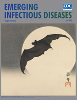
“On the Bat’s Back I Do Fly after Summer Merrily” [PDF - 2.14 MB - 3 pages]
| EID | Breedlove B. “On the Bat’s Back I Do Fly after Summer Merrily”. Emerg Infect Dis. 2023;29(7):1499-1501. https://doi.org/10.3201/eid2907.ac2907 |
|---|---|
| AMA | Breedlove B. “On the Bat’s Back I Do Fly after Summer Merrily”. Emerging Infectious Diseases. 2023;29(7):1499-1501. doi:10.3201/eid2907.ac2907. |
| APA | Breedlove, B. (2023). “On the Bat’s Back I Do Fly after Summer Merrily”. Emerging Infectious Diseases, 29(7), 1499-1501. https://doi.org/10.3201/eid2907.ac2907. |


