Volume 9, Number 3—March 2003
Synopsis
Electron Microscopy for Rapid Diagnosis of Emerging Infectious Agents1
Paul R. Hazelton* and Hans R. Gelderblom†
Figure 7
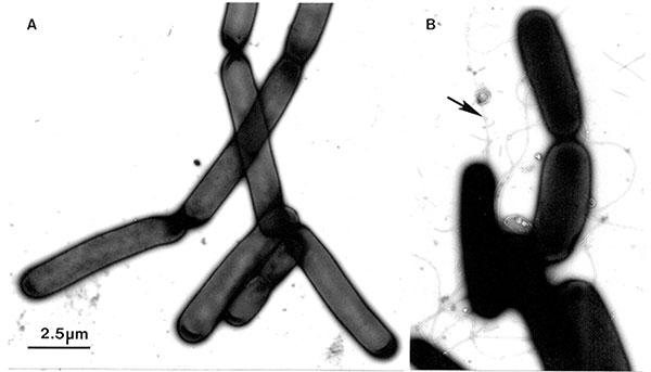
Figure 7. AA colony of Bacillus anthracis was suspended, inactivated, and negatively contrasted with aqueous uranyl acetate, as described for Figure 4The microorganisms, which grow in long chains, do not have flagellaBThe ubiquitous Bsubtilis may also grow as long chainsHowever, in contrast to Banthracis, the Bsubtilis cells show distinct flagella (arrow)Bar = 2.5 μm.
Page created: December 07, 2010
Page updated: December 07, 2010
Page reviewed: December 07, 2010
The conclusions, findings, and opinions expressed by authors contributing to this journal do not necessarily reflect the official position of the U.S. Department of Health and Human Services, the Public Health Service, the Centers for Disease Control and Prevention, or the authors' affiliated institutions. Use of trade names is for identification only and does not imply endorsement by any of the groups named above.