Synopses
Poxvirus Viability and Signatures in Historical Relics
Although it has been >30 years since the eradication of smallpox, the unearthing of well-preserved tissue material in which the virus may reside has called into question the viability of variola virus decades or centuries after its original occurrence. Experimental data to address the long-term stability and viability of the virus are limited. There are several instances of well-preserved corpses and tissues that have been examined for poxvirus viability and viral DNA. These historical specimens cause concern for potential exposures, and each situation should be approached cautiously and independently with the available information. Nevertheless, these specimens provide information on the history of a major disease and vaccination against it.
| EID | McCollum AM, Li Y, Wilkins K, Karem KL, Davidson WB, Paddock CD, et al. Poxvirus Viability and Signatures in Historical Relics. Emerg Infect Dis. 2014;20(2):177-184. https://doi.org/10.3201/eid2002.131098 |
|---|---|
| AMA | McCollum AM, Li Y, Wilkins K, et al. Poxvirus Viability and Signatures in Historical Relics. Emerging Infectious Diseases. 2014;20(2):177-184. doi:10.3201/eid2002.131098. |
| APA | McCollum, A. M., Li, Y., Wilkins, K., Karem, K. L., Davidson, W. B., Paddock, C. D....Damon, I. K. (2014). Poxvirus Viability and Signatures in Historical Relics. Emerging Infectious Diseases, 20(2), 177-184. https://doi.org/10.3201/eid2002.131098. |
Anncaliia algerae Microsporidial Myositis
The insect microsporidian Anncaliia algerae was first described in 2004 as a cause of fatal myositis in an immunosuppressed person from Pennsylvania, USA. Two cases were subsequently reported, and we detail 2 additional cases, including the only nonfatal case. We reviewed all 5 case histories with respect to clinical characteristics, diagnosis, and management and summarized organism life cycle and epidemiology. Before infection, all case-patients were using immunosuppressive medications for rheumatoid arthritis or solid-organ transplantation. Four of the 5 case-patients were from Australia. All diagnoses were confirmed by skeletal muscle biopsy; however, peripheral nerves and other tissues may be infected. The surviving patient received albendazole and had a reduction of immunosuppressive medications and measures to prevent complications. Although insects are the natural hosts for A. algerae, human contact with water contaminated by spores may be a mode of transmission. A. algerae has emerged as a cause of myositis, particularly in coastal Australia.
| EID | Watts MR, Chan R, Cheong E, Brammah S, Clezy KR, Tong C, et al. Anncaliia algerae Microsporidial Myositis. Emerg Infect Dis. 2014;20(2):185-191. https://doi.org/10.3201/eid2002.131126 |
|---|---|
| AMA | Watts MR, Chan R, Cheong E, et al. Anncaliia algerae Microsporidial Myositis. Emerging Infectious Diseases. 2014;20(2):185-191. doi:10.3201/eid2002.131126. |
| APA | Watts, M. R., Chan, R., Cheong, E., Brammah, S., Clezy, K. R., Tong, C....Reddel, S. W. (2014). Anncaliia algerae Microsporidial Myositis. Emerging Infectious Diseases, 20(2), 185-191. https://doi.org/10.3201/eid2002.131126. |
Research
Genomic Variability of Monkeypox Virus among Humans, Democratic Republic of the Congo
Monkeypox virus is a zoonotic virus endemic to Central Africa. Although active disease surveillance has assessed monkeypox disease prevalence and geographic range, information about virus diversity is lacking. We therefore assessed genome diversity of viruses in 60 samples obtained from humans with primary and secondary cases of infection from 2005 through 2007. We detected 4 distinct lineages and a deletion that resulted in gene loss in 10 (16.7%) samples and that seemed to correlate with human-to-human transmission (p = 0.0544). The data suggest a high frequency of spillover events from the pool of viruses in nonhuman animals, active selection through genomic destabilization and gene loss, and increased disease transmissibility and severity. The potential for accelerated adaptation to humans should be monitored through improved surveillance.
| EID | Kugelman JR, Johnston SC, Mulembakani PM, Kisalu N, Lee MS, Koroleva G, et al. Genomic Variability of Monkeypox Virus among Humans, Democratic Republic of the Congo. Emerg Infect Dis. 2014;20(2):232-239. https://doi.org/10.3201/eid2002.130118 |
|---|---|
| AMA | Kugelman JR, Johnston SC, Mulembakani PM, et al. Genomic Variability of Monkeypox Virus among Humans, Democratic Republic of the Congo. Emerging Infectious Diseases. 2014;20(2):232-239. doi:10.3201/eid2002.130118. |
| APA | Kugelman, J. R., Johnston, S. C., Mulembakani, P. M., Kisalu, N., Lee, M. S., Koroleva, G....Rimoin, A. W. (2014). Genomic Variability of Monkeypox Virus among Humans, Democratic Republic of the Congo. Emerging Infectious Diseases, 20(2), 232-239. https://doi.org/10.3201/eid2002.130118. |
Human Antibody Responses to Avian Influenza A(H7N9) Virus, 2013
Understanding host antibody response is crucial for predicting disease severity and for vaccine development. We investigated antibody responses against influenza A(H7N9) virus in 48 serum samples from 21 patients, including paired samples from 15 patients. IgG against subtype H7 and neutralizing antibodies (NAbs) were not detected in acute-phase samples, but ELISA geometric mean titers increased in convalescent-phase samples; NAb titers were 20–80 (geometric mean titer 40). Avidity to IgG against subtype H7 was significantly lower than that against H1 and H3. IgG against H3 was boosted after infection with influenza A(H7N9) virus, and its level in acute-phase samples correlated with that against H7 in convalescent-phase samples. A correlation was also found between hemagglutinin inhibition and NAb titers and between hemagglutinin inhibition and IgG titers against H7. Because of the relatively weak protective antibody response to influenza A(H7N9), multiple vaccinations might be needed to achieve protective immunity.
| EID | Guo L, Zhang X, Ren L, Yu X, Chen L, Zhou H, et al. Human Antibody Responses to Avian Influenza A(H7N9) Virus, 2013. Emerg Infect Dis. 2014;20(2):192-200. https://doi.org/10.3201/eid2002.131094 |
|---|---|
| AMA | Guo L, Zhang X, Ren L, et al. Human Antibody Responses to Avian Influenza A(H7N9) Virus, 2013. Emerging Infectious Diseases. 2014;20(2):192-200. doi:10.3201/eid2002.131094. |
| APA | Guo, L., Zhang, X., Ren, L., Yu, X., Chen, L., Zhou, H....Wang, J. (2014). Human Antibody Responses to Avian Influenza A(H7N9) Virus, 2013. Emerging Infectious Diseases, 20(2), 192-200. https://doi.org/10.3201/eid2002.131094. |
Seven-Valent Pneumococcal Conjugate Vaccine and Nasopharyngeal Microbiota in Healthy Children
Seven-valent pneumococcal conjugate vaccine (PCV-7) is effective against vaccine serotype disease and carriage. Nevertheless, shifts in colonization and disease toward nonvaccine serotypes and other potential pathogens have been described. To understand the extent of these shifts, we analyzed nasopharyngeal microbial profiles of 97 PCV-7–vaccinated infants and 103 control infants participating in a randomized controlled trial in the Netherlands. PCV-7 immunization resulted in a temporary shift in microbial community composition and increased bacterial diversity. Immunization also resulted in decreased presence of the pneumococcal vaccine serotype and an increase in the relative abundance and presence of nonpneumococcal streptococci and anaerobic bacteria. Furthermore, the abundance of Haemophilus and Staphylococcus bacteria in vaccinees was increased over that in controls. This study illustrates the much broader effect of vaccination with PCV-7 on the microbial community than currently assumed, and highlights the need for careful monitoring when implementing vaccines directed against common colonizers.
| EID | Biesbroek G, Wang X, Keijser B, Eijkemans R, Trzciński K, Rots NY, et al. Seven-Valent Pneumococcal Conjugate Vaccine and Nasopharyngeal Microbiota in Healthy Children. Emerg Infect Dis. 2014;20(2):201-210. https://doi.org/10.3201/eid2002.131220 |
|---|---|
| AMA | Biesbroek G, Wang X, Keijser B, et al. Seven-Valent Pneumococcal Conjugate Vaccine and Nasopharyngeal Microbiota in Healthy Children. Emerging Infectious Diseases. 2014;20(2):201-210. doi:10.3201/eid2002.131220. |
| APA | Biesbroek, G., Wang, X., Keijser, B., Eijkemans, R., Trzciński, K., Rots, N. Y....Bogaert, D. (2014). Seven-Valent Pneumococcal Conjugate Vaccine and Nasopharyngeal Microbiota in Healthy Children. Emerging Infectious Diseases, 20(2), 201-210. https://doi.org/10.3201/eid2002.131220. |
Fungal Endophthalmitis Associated with Compounded Products
Fungal endophthalmitis is a rare but serious infection. In March 2012, several cases of probable and laboratory-confirmed fungal endophthalmitis occurring after invasive ocular procedures were reported nationwide. We identified 47 cases in 9 states: 21 patients had been exposed to the intraocular dye Brilliant Blue G (BBG) during retinal surgery, and the other 26 had received an intravitreal injection containing triamcinolone acetonide. Both drugs were produced by Franck’s Compounding Lab (Ocala, FL, USA). Fusarium incarnatum-equiseti species complex mold was identified in specimens from BBG-exposed case-patients and an unopened BBG vial. Bipolaris hawaiiensis mold was identified in specimens from triamcinolone-exposed case-patients. Exposure to either product was the only factor associated with case status. Of 40 case-patients for whom data were available, 39 (98%) lost vision. These concurrent outbreaks, associated with 1 compounding pharmacy, resulted in a product recall. Ensuring safety and integrity of compounded medications is critical for preventing further outbreaks associated with compounded products.
| EID | Mikosz CA, Smith RM, Kim M, Tyson C, Lee EH, Adams E, et al. Fungal Endophthalmitis Associated with Compounded Products. Emerg Infect Dis. 2014;20(2):248-256. https://doi.org/10.3201/eid2002.131257 |
|---|---|
| AMA | Mikosz CA, Smith RM, Kim M, et al. Fungal Endophthalmitis Associated with Compounded Products. Emerging Infectious Diseases. 2014;20(2):248-256. doi:10.3201/eid2002.131257. |
| APA | Mikosz, C. A., Smith, R. M., Kim, M., Tyson, C., Lee, E. H., Adams, E....Park, B. J. (2014). Fungal Endophthalmitis Associated with Compounded Products. Emerging Infectious Diseases, 20(2), 248-256. https://doi.org/10.3201/eid2002.131257. |
Monitoring Human Babesiosis Emergence through Vector Surveillance New England, USA
Human babesiosis is an emerging tick-borne disease caused by the intraerythrocytic protozoan Babesia microti. Its geographic distribution is more limited than that of Lyme disease, despite sharing the same tick vector and reservoir hosts. The geographic range of babesiosis is expanding, but knowledge of its range is incomplete and relies exclusively on reports of human cases. We evaluated the utility of tick-based surveillance for monitoring disease expansion by comparing the ratios of the 2 infections in humans and ticks in areas with varying B. microti endemicity. We found a close association between human disease and tick infection ratios in long-established babesiosis-endemic areas but a lower than expected incidence of human babesiosis on the basis of tick infection rates in new disease-endemic areas. This finding suggests that babesiosis at emerging sites is underreported. Vector-based surveillance can provide an early warning system for the emergence of human babesiosis.
| EID | Diuk-Wasser MA, Liu Y, Steeves TK, Folsom-O’Keefe C, Dardick KR, Lepore T, et al. Monitoring Human Babesiosis Emergence through Vector Surveillance New England, USA. Emerg Infect Dis. 2014;20(2):225-231. https://doi.org/10.3201/eid2002.130644 |
|---|---|
| AMA | Diuk-Wasser MA, Liu Y, Steeves TK, et al. Monitoring Human Babesiosis Emergence through Vector Surveillance New England, USA. Emerging Infectious Diseases. 2014;20(2):225-231. doi:10.3201/eid2002.130644. |
| APA | Diuk-Wasser, M. A., Liu, Y., Steeves, T. K., Folsom-O’Keefe, C., Dardick, K. R., Lepore, T....Krause, P. J. (2014). Monitoring Human Babesiosis Emergence through Vector Surveillance New England, USA. Emerging Infectious Diseases, 20(2), 225-231. https://doi.org/10.3201/eid2002.130644. |
Lymphocytic Choriomeningitis Virus in Employees and Mice at Multipremises Feeder-Rodent Operation, United States, 2012
We investigated the extent of lymphocytic choriomeningitis virus (LCMV) infection in employees and rodents at 3 commercial breeding facilities. Of 97 employees tested, 31 (32%) had IgM and/or IgG to LCMV, and aseptic meningitis was diagnosed in 4 employees. Of 1,820 rodents tested in 1 facility, 382 (21%) mice (Mus musculus) had detectable IgG, and 13 (0.7%) were positive by reverse transcription PCR; LCMV was isolated from 8. Rats (Rattus norvegicus) were not found to be infected. S-segment RNA sequence was similar to strains previously isolated in North America. Contact by wild mice with colony mice was the likely source for LCMV, and shipments of infected mice among facilities spread the infection. The breeding colonies were depopulated to prevent further human infections. Future outbreaks can be prevented with monitoring and management, and employees should be made aware of LCMV risks and prevention.
| EID | Knust B, Ströher U, Edison L, Albariño CG, Lovejoy J, Armeanu E, et al. Lymphocytic Choriomeningitis Virus in Employees and Mice at Multipremises Feeder-Rodent Operation, United States, 2012. Emerg Infect Dis. 2014;20(2):240-247. https://doi.org/10.3201/eid2002.130860 |
|---|---|
| AMA | Knust B, Ströher U, Edison L, et al. Lymphocytic Choriomeningitis Virus in Employees and Mice at Multipremises Feeder-Rodent Operation, United States, 2012. Emerging Infectious Diseases. 2014;20(2):240-247. doi:10.3201/eid2002.130860. |
| APA | Knust, B., Ströher, U., Edison, L., Albariño, C. G., Lovejoy, J., Armeanu, E....Rollin, P. E. (2014). Lymphocytic Choriomeningitis Virus in Employees and Mice at Multipremises Feeder-Rodent Operation, United States, 2012. Emerging Infectious Diseases, 20(2), 240-247. https://doi.org/10.3201/eid2002.130860. |
Subtyping Cryptosporidium ubiquitum,a Zoonotic Pathogen Emerging in Humans
Cryptosporidium ubiquitum is an emerging zoonotic pathogen. In the past, it was not possible to identify an association between cases of human and animal infection. We conducted a genomic survey of the species, developed a subtyping tool targeting the 60-kDa glycoprotein (gp60) gene, and identified 6 subtype families (XIIa–XIIf) of C. ubiquitum. Host adaptation was apparent at the gp60 locus; subtype XIIa was found in ruminants worldwide, subtype families XIIb–XIId were found in rodents in the United States, and XIIe and XIIf were found in rodents in the Slovak Republic. Humans in the United States were infected with isolates of subtypes XIIb–XIId, whereas those in other areas were infected primarily with subtype XIIa isolates. In addition, subtype families XIIb and XIId were detected in drinking source water in the United States. Contact with C. ubiquitum–infected sheep and drinking water contaminated by infected wildlife could be sources of human infections.
| EID | Li N, Xiao L, Alderisio K, Elwin K, Cebelinski E, Chalmers R, et al. Subtyping Cryptosporidium ubiquitum,a Zoonotic Pathogen Emerging in Humans. Emerg Infect Dis. 2014;20(2):217-224. https://doi.org/10.3201/eid2002.121797 |
|---|---|
| AMA | Li N, Xiao L, Alderisio K, et al. Subtyping Cryptosporidium ubiquitum,a Zoonotic Pathogen Emerging in Humans. Emerging Infectious Diseases. 2014;20(2):217-224. doi:10.3201/eid2002.121797. |
| APA | Li, N., Xiao, L., Alderisio, K., Elwin, K., Cebelinski, E., Chalmers, R....Feng, Y. (2014). Subtyping Cryptosporidium ubiquitum,a Zoonotic Pathogen Emerging in Humans. Emerging Infectious Diseases, 20(2), 217-224. https://doi.org/10.3201/eid2002.121797. |
Novel Paramyxovirus Associated with Severe Acute Febrile Disease, South Sudan and Uganda, 2012
In 2012, a female wildlife biologist experienced fever, malaise, headache, generalized myalgia and arthralgia, neck stiffness, and a sore throat shortly after returning to the United States from a 6-week field expedition to South Sudan and Uganda. She was hospitalized, after which a maculopapular rash developed and became confluent. When the patient was discharged from the hospital on day 14, arthralgia and myalgia had improved, oropharynx ulcerations had healed, the rash had resolved without desquamation, and blood counts and hepatic enzyme levels were returning to reference levels. After several known suspect pathogens were ruled out as the cause of her illness, deep sequencing and metagenomics analysis revealed a novel paramyxovirus related to rubula-like viruses isolated from fruit bats.
| EID | Albariño CG, Foltzer M, Towner JS, Rowe LA, Campbell S, Jaramillo CM, et al. Novel Paramyxovirus Associated with Severe Acute Febrile Disease, South Sudan and Uganda, 2012. Emerg Infect Dis. 2014;20(2):211-216. https://doi.org/10.3201/eid2002.131620 |
|---|---|
| AMA | Albariño CG, Foltzer M, Towner JS, et al. Novel Paramyxovirus Associated with Severe Acute Febrile Disease, South Sudan and Uganda, 2012. Emerging Infectious Diseases. 2014;20(2):211-216. doi:10.3201/eid2002.131620. |
| APA | Albariño, C. G., Foltzer, M., Towner, J. S., Rowe, L. A., Campbell, S., Jaramillo, C. M....Ströher, U. (2014). Novel Paramyxovirus Associated with Severe Acute Febrile Disease, South Sudan and Uganda, 2012. Emerging Infectious Diseases, 20(2), 211-216. https://doi.org/10.3201/eid2002.131620. |
Dispatches
Melioidosis Caused by Burkholderia pseudomallei in Drinking Water, Thailand, 2012
We identified 10 patients in Thailand with culture-confirmed melioidosis who had Burkholderia pseudomallei isolated from their drinking water. The multilocus sequence type of B. pseudomallei from clinical specimens and water samples were identical for 2 patients. This finding suggests that drinking water is a preventable source of B. pseudomallei infection.
| EID | Limmathurotsakul D, Wongsuvan G, Aanensen D, Ngamwilai S, Saiprom N, Rongkard P, et al. Melioidosis Caused by Burkholderia pseudomallei in Drinking Water, Thailand, 2012. Emerg Infect Dis. 2014;20(2):265-268. https://doi.org/10.3201/eid2002.121891 |
|---|---|
| AMA | Limmathurotsakul D, Wongsuvan G, Aanensen D, et al. Melioidosis Caused by Burkholderia pseudomallei in Drinking Water, Thailand, 2012. Emerging Infectious Diseases. 2014;20(2):265-268. doi:10.3201/eid2002.121891. |
| APA | Limmathurotsakul, D., Wongsuvan, G., Aanensen, D., Ngamwilai, S., Saiprom, N., Rongkard, P....Peacock, S. J. (2014). Melioidosis Caused by Burkholderia pseudomallei in Drinking Water, Thailand, 2012. Emerging Infectious Diseases, 20(2), 265-268. https://doi.org/10.3201/eid2002.121891. |
Genetic Characterization of Coronaviruses from Domestic Ferrets, Japan
We detected ferret coronaviruses in 44 (55.7%) of 79 pet ferrets tested in Japan and classified the viruses into 2 genotypes on the basis of genotype-specific PCR. Our results show that 2 ferret coronaviruses that cause feline infectious peritonitis–like disease and epizootic catarrhal enteritis are enzootic among ferrets in Japan.
| EID | Terada Y, Minami S, Noguchi K, Mahmoud H, Shimoda H, Mochizuki M, et al. Genetic Characterization of Coronaviruses from Domestic Ferrets, Japan. Emerg Infect Dis. 2014;20(2):284-287. https://doi.org/10.3201/eid2002.130543 |
|---|---|
| AMA | Terada Y, Minami S, Noguchi K, et al. Genetic Characterization of Coronaviruses from Domestic Ferrets, Japan. Emerging Infectious Diseases. 2014;20(2):284-287. doi:10.3201/eid2002.130543. |
| APA | Terada, Y., Minami, S., Noguchi, K., Mahmoud, H., Shimoda, H., Mochizuki, M....Maeda, K. (2014). Genetic Characterization of Coronaviruses from Domestic Ferrets, Japan. Emerging Infectious Diseases, 20(2), 284-287. https://doi.org/10.3201/eid2002.130543. |
Reemergence of Rift Valley Fever, Mauritania, 2010
A Rift Valley fever (RVF) outbreak in humans and animals occurred in Mauritania in 2010. Thirty cases of RVF in humans and 3 deaths were identified. RVFV isolates were recovered from humans, camels, sheep, goats, and Culex antennatus mosquitoes. Phylogenetic analysis of isolates indicated a virus origin from western Africa.
| EID | Faye O, Ba H, Ba Y, Freire C, Faye O, Ndiaye O, et al. Reemergence of Rift Valley Fever, Mauritania, 2010. Emerg Infect Dis. 2014;20(2):300-303. https://doi.org/10.3201/eid2002.130996 |
|---|---|
| AMA | Faye O, Ba H, Ba Y, et al. Reemergence of Rift Valley Fever, Mauritania, 2010. Emerging Infectious Diseases. 2014;20(2):300-303. doi:10.3201/eid2002.130996. |
| APA | Faye, O., Ba, H., Ba, Y., Freire, C., Faye, O., Ndiaye, O....Sall, A. A. (2014). Reemergence of Rift Valley Fever, Mauritania, 2010. Emerging Infectious Diseases, 20(2), 300-303. https://doi.org/10.3201/eid2002.130996. |
Rift Valley Fever Outbreak, Southern Mauritania, 2012
After a period of heavy rainfall, an outbreak of Rift Valley fever occurred in southern Mauritania during September–November 2012. A total of 41 human cases were confirmed, including 13 deaths, and 12 Rift Valley fever virus strains were isolated. Moudjeria and Temchecket Departments were the most affected areas.
| EID | Sow A, Faye O, Ba Y, Ba H, Diallo D, Faye O, et al. Rift Valley Fever Outbreak, Southern Mauritania, 2012. Emerg Infect Dis. 2014;20(2):296-299. https://doi.org/10.3201/eid2002.131000 |
|---|---|
| AMA | Sow A, Faye O, Ba Y, et al. Rift Valley Fever Outbreak, Southern Mauritania, 2012. Emerging Infectious Diseases. 2014;20(2):296-299. doi:10.3201/eid2002.131000. |
| APA | Sow, A., Faye, O., Ba, Y., Ba, H., Diallo, D., Faye, O....Sall, A. (2014). Rift Valley Fever Outbreak, Southern Mauritania, 2012. Emerging Infectious Diseases, 20(2), 296-299. https://doi.org/10.3201/eid2002.131000. |
Co-circulation of West Nile Virus Variants, Arizona, USA, 2010
Molecular analysis of West Nile virus (WNV) isolates obtained during a 2010 outbreak in Maricopa County, Arizona, USA, demonstrated co-circulation of 3 distinct genetic variants, including strains with novel envelope protein mutations. These results highlight the continuing evolution of WNV in North America and the current complexity of WNV dispersal and transmission.
| EID | Plante JA, Burkhalter KL, Mann BR, Godsey MS, Mutebi J, Beasley D. Co-circulation of West Nile Virus Variants, Arizona, USA, 2010. Emerg Infect Dis. 2014;20(2):272-275. https://doi.org/10.3201/eid2002.131008 |
|---|---|
| AMA | Plante JA, Burkhalter KL, Mann BR, et al. Co-circulation of West Nile Virus Variants, Arizona, USA, 2010. Emerging Infectious Diseases. 2014;20(2):272-275. doi:10.3201/eid2002.131008. |
| APA | Plante, J. A., Burkhalter, K. L., Mann, B. R., Godsey, M. S., Mutebi, J., & Beasley, D. (2014). Co-circulation of West Nile Virus Variants, Arizona, USA, 2010. Emerging Infectious Diseases, 20(2), 272-275. https://doi.org/10.3201/eid2002.131008. |
Andes Hantavirus Variant in Rodents, Southern Amazon Basin, Peru
We investigated hantaviruses in rodents in the southern Amazon Basin of Peru and identified an Andes virus variant from Neacomys spinosus mice. This finding extends the known range of this virus in South America and the range of recognized hantaviruses in Peru. Further studies of the epizoology of hantaviruses in this region are warranted.
| EID | Razuri H, Tokarz R, Ghersi BM, Salmon-Mulanovich G, Guezala M, Albujar C, et al. Andes Hantavirus Variant in Rodents, Southern Amazon Basin, Peru. Emerg Infect Dis. 2014;20(2):257-260. https://doi.org/10.3201/eid2002.131418 |
|---|---|
| AMA | Razuri H, Tokarz R, Ghersi BM, et al. Andes Hantavirus Variant in Rodents, Southern Amazon Basin, Peru. Emerging Infectious Diseases. 2014;20(2):257-260. doi:10.3201/eid2002.131418. |
| APA | Razuri, H., Tokarz, R., Ghersi, B. M., Salmon-Mulanovich, G., Guezala, M., Albujar, C....Montgomery, J. M. (2014). Andes Hantavirus Variant in Rodents, Southern Amazon Basin, Peru. Emerging Infectious Diseases, 20(2), 257-260. https://doi.org/10.3201/eid2002.131418. |
Crimean-Congo Hemorrhagic Fever Virus, Greece
Seroprevalence of Crimean-Congo hemorrhagic fever virus (CCHFV) is high in some regions of Greece, but only 1 case of disease has been reported. We used 4 methods to test 118 serum samples that were positive for CCHFV IgG by commercial ELISA and confirmed the positive results. A nonpathogenic or low-pathogenicity strain may be circulating.
| EID | Papa A, Sidira P, Larichev V, Gavrilova L, Kuzmina K, Mousavi-Jazi M, et al. Crimean-Congo Hemorrhagic Fever Virus, Greece. Emerg Infect Dis. 2014;20(2):288-290. https://doi.org/10.3201/eid2002.130690 |
|---|---|
| AMA | Papa A, Sidira P, Larichev V, et al. Crimean-Congo Hemorrhagic Fever Virus, Greece. Emerging Infectious Diseases. 2014;20(2):288-290. doi:10.3201/eid2002.130690. |
| APA | Papa, A., Sidira, P., Larichev, V., Gavrilova, L., Kuzmina, K., Mousavi-Jazi, M....Nichol, S. (2014). Crimean-Congo Hemorrhagic Fever Virus, Greece. Emerging Infectious Diseases, 20(2), 288-290. https://doi.org/10.3201/eid2002.130690. |
Lethal Factor and Anti-Protective Antigen IgG Levels Associated with Inhalation Anthrax, Minnesota, USA
Bacillus anthracis was identified in a 61-year-old man hospitalized in Minnesota, USA. Cooperation between the hospital and the state health agency enhanced prompt identification of the pathogen. Treatment comprising antimicrobial drugs, anthrax immune globulin, and pleural drainage led to full recovery; however, the role of passive immunization in anthrax treatment requires further evaluation.
| EID | Sprenkle MD, Griffith J, Marinelli W, Boyer AE, Quinn CP, Pesik NT, et al. Lethal Factor and Anti-Protective Antigen IgG Levels Associated with Inhalation Anthrax, Minnesota, USA. Emerg Infect Dis. 2014;20(2):310-314. https://doi.org/10.3201/eid2002.130245 |
|---|---|
| AMA | Sprenkle MD, Griffith J, Marinelli W, et al. Lethal Factor and Anti-Protective Antigen IgG Levels Associated with Inhalation Anthrax, Minnesota, USA. Emerging Infectious Diseases. 2014;20(2):310-314. doi:10.3201/eid2002.130245. |
| APA | Sprenkle, M. D., Griffith, J., Marinelli, W., Boyer, A. E., Quinn, C. P., Pesik, N. T....Blaney, D. D. (2014). Lethal Factor and Anti-Protective Antigen IgG Levels Associated with Inhalation Anthrax, Minnesota, USA. Emerging Infectious Diseases, 20(2), 310-314. https://doi.org/10.3201/eid2002.130245. |
Replicative Capacity of MERS Coronavirus in Livestock Cell Lines
Replicative capacity of Middle East respiratory syndrome coronavirus (MERS-CoV) was assessed in cell lines derived from livestock and peridomestic small mammals on the Arabian Peninsula. Only cell lines originating from goats and camels showed efficient replication of MERS-CoV. These results provide direction in the search for the intermediate host of MERS-CoV.
| EID | Eckerle I, Corman VM, Müller MA, Lenk M, Ulrich RG, Drosten C. Replicative Capacity of MERS Coronavirus in Livestock Cell Lines. Emerg Infect Dis. 2014;20(2):276-279. https://doi.org/10.3201/eid2002.131182 |
|---|---|
| AMA | Eckerle I, Corman VM, Müller MA, et al. Replicative Capacity of MERS Coronavirus in Livestock Cell Lines. Emerging Infectious Diseases. 2014;20(2):276-279. doi:10.3201/eid2002.131182. |
| APA | Eckerle, I., Corman, V. M., Müller, M. A., Lenk, M., Ulrich, R. G., & Drosten, C. (2014). Replicative Capacity of MERS Coronavirus in Livestock Cell Lines. Emerging Infectious Diseases, 20(2), 276-279. https://doi.org/10.3201/eid2002.131182. |
Investigation of Inhalation Anthrax Case, United States
Inhalation anthrax occurred in a man who vacationed in 4 US states where anthrax is enzootic. Despite an extensive multi-agency investigation, the specific source was not detected, and no additional related human or animal cases were found. Although rare, inhalation anthrax can occur naturally in the United States.
| EID | Griffith J, Blaney D, Shadomy S, Lehman M, Pesik N, Tostenson S, et al. Investigation of Inhalation Anthrax Case, United States. Emerg Infect Dis. 2014;20(2):280-283. https://doi.org/10.3201/eid2002.130021 |
|---|---|
| AMA | Griffith J, Blaney D, Shadomy S, et al. Investigation of Inhalation Anthrax Case, United States. Emerging Infectious Diseases. 2014;20(2):280-283. doi:10.3201/eid2002.130021. |
| APA | Griffith, J., Blaney, D., Shadomy, S., Lehman, M., Pesik, N., Tostenson, S....Lynfield, R. (2014). Investigation of Inhalation Anthrax Case, United States. Emerging Infectious Diseases, 20(2), 280-283. https://doi.org/10.3201/eid2002.130021. |
Burkholderia pseudomallei Isolates in 2 Pet Iguanas, California, USA
Burkholderia pseudomallei, the causative agent of melioidosis, was isolated from abscesses of 2 pet green iguanas in California, USA. The international trade in iguanas may contribute to importation of this pathogen into countries where it is not endemic and put persons exposed to these animals at risk for infection.
| EID | Zehnder AM, Hawkins MG, Koski MA, Lifland B, Byrne BA, Swanson AA, et al. Burkholderia pseudomallei Isolates in 2 Pet Iguanas, California, USA. Emerg Infect Dis. 2014;20(2):304-306. https://doi.org/10.3201/eid2002.131314 |
|---|---|
| AMA | Zehnder AM, Hawkins MG, Koski MA, et al. Burkholderia pseudomallei Isolates in 2 Pet Iguanas, California, USA. Emerging Infectious Diseases. 2014;20(2):304-306. doi:10.3201/eid2002.131314. |
| APA | Zehnder, A. M., Hawkins, M. G., Koski, M. A., Lifland, B., Byrne, B. A., Swanson, A. A....Beeler, E. S. (2014). Burkholderia pseudomallei Isolates in 2 Pet Iguanas, California, USA. Emerging Infectious Diseases, 20(2), 304-306. https://doi.org/10.3201/eid2002.131314. |
Molecular Detection of Diphyllobothrium nihonkaiense in Humans, China
The cause of diphyllobothriosis in 5 persons in Harbin and Shanghai, China, during 2008–2011, initially attributed to the tapeworm Diphyllobothrium latum, was confirmed as D. nihonkaiense by using molecular analysis of expelled proglottids. The use of morphologic characteristics alone to identify this organism was inadequate and led to misidentification of the species.
| EID | Chen S, Ai L, Zhang Y, Chen J, Zhang W, Li Y, et al. Molecular Detection of Diphyllobothrium nihonkaiense in Humans, China. Emerg Infect Dis. 2014;20(2):315-318. https://doi.org/10.3201/eid2002.121889 |
|---|---|
| AMA | Chen S, Ai L, Zhang Y, et al. Molecular Detection of Diphyllobothrium nihonkaiense in Humans, China. Emerging Infectious Diseases. 2014;20(2):315-318. doi:10.3201/eid2002.121889. |
| APA | Chen, S., Ai, L., Zhang, Y., Chen, J., Zhang, W., Li, Y....Yamasaki, H. (2014). Molecular Detection of Diphyllobothrium nihonkaiense in Humans, China. Emerging Infectious Diseases, 20(2), 315-318. https://doi.org/10.3201/eid2002.121889. |
Human Cutaneous Anthrax, Georgia 2010–2012
We assessed the occurrence of human cutaneous anthrax in Georgia during 2010–-2012 by examining demographic and spatial characteristics of reported cases. Reporting increased substantially, as did clustering of cases near urban centers. Control efforts, including education about anthrax and livestock vaccination, can be directed at areas of high risk.
| EID | Kracalik I, Malania L, Tsertsvadze N, Manvelyan J, Bakanidze L, Imnadze P, et al. Human Cutaneous Anthrax, Georgia 2010–2012. Emerg Infect Dis. 2014;20(2):261-264. https://doi.org/10.3201/eid2002.130522 |
|---|---|
| AMA | Kracalik I, Malania L, Tsertsvadze N, et al. Human Cutaneous Anthrax, Georgia 2010–2012. Emerging Infectious Diseases. 2014;20(2):261-264. doi:10.3201/eid2002.130522. |
| APA | Kracalik, I., Malania, L., Tsertsvadze, N., Manvelyan, J., Bakanidze, L., Imnadze, P....Blackburn, J. K. (2014). Human Cutaneous Anthrax, Georgia 2010–2012. Emerging Infectious Diseases, 20(2), 261-264. https://doi.org/10.3201/eid2002.130522. |
Fatal Systemic Morbillivirus Infection in Bottlenose Dolphin, Canary Islands, Spain
A systemic morbillivirus infection was diagnosed postmortem in a juvenile bottlenose dolphin stranded in the eastern North Atlantic Ocean in 2005. Sequence analysis of a conserved fragment of the morbillivirus phosphoprotein gene indicated that the virus is closely related to dolphin morbillivirus recently reported in striped dolphins in the Mediterranean Sea.
| EID | Sierra E, Zucca D, Arbelo M, García-Álvarez N, Andrada M, Déniz S, et al. Fatal Systemic Morbillivirus Infection in Bottlenose Dolphin, Canary Islands, Spain. Emerg Infect Dis. 2014;20(2):269-271. https://doi.org/10.3201/eid2002.131463 |
|---|---|
| AMA | Sierra E, Zucca D, Arbelo M, et al. Fatal Systemic Morbillivirus Infection in Bottlenose Dolphin, Canary Islands, Spain. Emerging Infectious Diseases. 2014;20(2):269-271. doi:10.3201/eid2002.131463. |
| APA | Sierra, E., Zucca, D., Arbelo, M., García-Álvarez, N., Andrada, M., Déniz, S....Fernández, A. (2014). Fatal Systemic Morbillivirus Infection in Bottlenose Dolphin, Canary Islands, Spain. Emerging Infectious Diseases, 20(2), 269-271. https://doi.org/10.3201/eid2002.131463. |
Congenital Rubella Syndrome in Child of Woman without Known Risk Factors, New Jersey, USA
We report a case of congenital rubella syndrome in a child born to a vaccinated New Jersey woman who had not traveled internationally. Although rubella and congenital rubella syndrome have been eliminated from the United States, clinicians should remain vigilant and immediately notify public health authorities when either is suspected.
| EID | Pitts SI, Wallace GS, Montana B, Handschur EF, Meislich D, Sampson AC, et al. Congenital Rubella Syndrome in Child of Woman without Known Risk Factors, New Jersey, USA. Emerg Infect Dis. 2014;20(2):307-309. https://doi.org/10.3201/eid2002.131233 |
|---|---|
| AMA | Pitts SI, Wallace GS, Montana B, et al. Congenital Rubella Syndrome in Child of Woman without Known Risk Factors, New Jersey, USA. Emerging Infectious Diseases. 2014;20(2):307-309. doi:10.3201/eid2002.131233. |
| APA | Pitts, S. I., Wallace, G. S., Montana, B., Handschur, E. F., Meislich, D., Sampson, A. C....Icenogle, J. P. (2014). Congenital Rubella Syndrome in Child of Woman without Known Risk Factors, New Jersey, USA. Emerging Infectious Diseases, 20(2), 307-309. https://doi.org/10.3201/eid2002.131233. |
Trace-Forward Investigation of Mice in Response to Lymphocytic Choriomeningitis Virus Outbreak
During follow-up of a 2012 US outbreak of lymphocytic choriomeningitis virus (LCMV), we conducted a trace-forward investigation. LCMV-infected feeder mice originating from a US rodent breeding facility had been distributed to >500 locations in 21 states. All mice from the facility were euthanized, and no additional persons tested positive for LCMV infection.
| EID | Edison L, Knust B, Petersen B, Gabel J, Manning C, Drenzek C, et al. Trace-Forward Investigation of Mice in Response to Lymphocytic Choriomeningitis Virus Outbreak. Emerg Infect Dis. 2014;20(2):291-295. https://doi.org/10.3201/eid2002.130861 |
|---|---|
| AMA | Edison L, Knust B, Petersen B, et al. Trace-Forward Investigation of Mice in Response to Lymphocytic Choriomeningitis Virus Outbreak. Emerging Infectious Diseases. 2014;20(2):291-295. doi:10.3201/eid2002.130861. |
| APA | Edison, L., Knust, B., Petersen, B., Gabel, J., Manning, C., Drenzek, C....Nichol, S. T. (2014). Trace-Forward Investigation of Mice in Response to Lymphocytic Choriomeningitis Virus Outbreak. Emerging Infectious Diseases, 20(2), 291-295. https://doi.org/10.3201/eid2002.130861. |
Commentaries
Low-Incidence, High-Consequence Pathogens
| EID | Belay ED, Monroe SS. Low-Incidence, High-Consequence Pathogens. Emerg Infect Dis. 2014;20(2):319-321. https://doi.org/10.3201/eid2002.131748 |
|---|---|
| AMA | Belay ED, Monroe SS. Low-Incidence, High-Consequence Pathogens. Emerging Infectious Diseases. 2014;20(2):319-321. doi:10.3201/eid2002.131748. |
| APA | Belay, E. D., & Monroe, S. S. (2014). Low-Incidence, High-Consequence Pathogens. Emerging Infectious Diseases, 20(2), 319-321. https://doi.org/10.3201/eid2002.131748. |
Letters
Laboratory-acquired Buffalopox Virus Infection, India
| EID | Riyesh T, Karuppusamy S, Bera BC, Barua S, Virmani N, Yadav S, et al. Laboratory-acquired Buffalopox Virus Infection, India. Emerg Infect Dis. 2014;20(2):324-326. https://doi.org/10.3201/eid2002.130358 |
|---|---|
| AMA | Riyesh T, Karuppusamy S, Bera BC, et al. Laboratory-acquired Buffalopox Virus Infection, India. Emerging Infectious Diseases. 2014;20(2):324-326. doi:10.3201/eid2002.130358. |
| APA | Riyesh, T., Karuppusamy, S., Bera, B. C., Barua, S., Virmani, N., Yadav, S....Singh, R. K. (2014). Laboratory-acquired Buffalopox Virus Infection, India. Emerging Infectious Diseases, 20(2), 324-326. https://doi.org/10.3201/eid2002.130358. |
Avian Influenza A(H7N9) Virus Infection in Pregnant Woman, China, 2013
| EID | Qi X, Cui L, Xu K, Wu B, Tang F, Bao C, et al. Avian Influenza A(H7N9) Virus Infection in Pregnant Woman, China, 2013. Emerg Infect Dis. 2014;20(2):332-333. https://doi.org/10.3201/eid2002.131109 |
|---|---|
| AMA | Qi X, Cui L, Xu K, et al. Avian Influenza A(H7N9) Virus Infection in Pregnant Woman, China, 2013. Emerging Infectious Diseases. 2014;20(2):332-333. doi:10.3201/eid2002.131109. |
| APA | Qi, X., Cui, L., Xu, K., Wu, B., Tang, F., Bao, C....Wang, H. (2014). Avian Influenza A(H7N9) Virus Infection in Pregnant Woman, China, 2013. Emerging Infectious Diseases, 20(2), 332-333. https://doi.org/10.3201/eid2002.131109. |
Stable Transmission of Dirofilaria repens Nematodes, Northern Germany
| EID | Czajka C, Becker N, Jöst H, Poppert S, Schmidt-Chanasit J, Krüger A, et al. Stable Transmission of Dirofilaria repens Nematodes, Northern Germany. Emerg Infect Dis. 2014;20(2):328-330. https://doi.org/10.3201/eid2002.131003 |
|---|---|
| AMA | Czajka C, Becker N, Jöst H, et al. Stable Transmission of Dirofilaria repens Nematodes, Northern Germany. Emerging Infectious Diseases. 2014;20(2):328-330. doi:10.3201/eid2002.131003. |
| APA | Czajka, C., Becker, N., Jöst, H., Poppert, S., Schmidt-Chanasit, J., Krüger, A....Tannich, E. (2014). Stable Transmission of Dirofilaria repens Nematodes, Northern Germany. Emerging Infectious Diseases, 20(2), 328-330. https://doi.org/10.3201/eid2002.131003. |
Peste des Petits Ruminants Virus, Mauritania
| EID | El Arbi A, El Mamy A, Salami H, Isselmou E, Kwiatek O, Libeau G, et al. Peste des Petits Ruminants Virus, Mauritania. Emerg Infect Dis. 2014;20(2):333-336. https://doi.org/10.3201/eid2002.131345 |
|---|---|
| AMA | El Arbi A, El Mamy A, Salami H, et al. Peste des Petits Ruminants Virus, Mauritania. Emerging Infectious Diseases. 2014;20(2):333-336. doi:10.3201/eid2002.131345. |
| APA | El Arbi, A., El Mamy, A., Salami, H., Isselmou, E., Kwiatek, O., Libeau, G....Lancelot, R. (2014). Peste des Petits Ruminants Virus, Mauritania. Emerging Infectious Diseases, 20(2), 333-336. https://doi.org/10.3201/eid2002.131345. |
Infectious Schmallenberg Virus from Bovine Semen, Germany
| EID | Schulz C, Wernike K, Beer M, Hoffmann B. Infectious Schmallenberg Virus from Bovine Semen, Germany. Emerg Infect Dis. 2014;20(2):337-339. https://doi.org/10.3201/eid2002.131436 |
|---|---|
| AMA | Schulz C, Wernike K, Beer M, et al. Infectious Schmallenberg Virus from Bovine Semen, Germany. Emerging Infectious Diseases. 2014;20(2):337-339. doi:10.3201/eid2002.131436. |
| APA | Schulz, C., Wernike, K., Beer, M., & Hoffmann, B. (2014). Infectious Schmallenberg Virus from Bovine Semen, Germany. Emerging Infectious Diseases, 20(2), 337-339. https://doi.org/10.3201/eid2002.131436. |
Novel Bunyavirus in Domestic and Captive Farmed Animals, Minnesota, USA
| EID | Nasci RS, Brault AC, Lambert AJ, Savage HM. Novel Bunyavirus in Domestic and Captive Farmed Animals, Minnesota, USA. Emerg Infect Dis. 2014;20(2):336. https://doi.org/10.3201/eid2002.131360 |
|---|---|
| AMA | Nasci RS, Brault AC, Lambert AJ, et al. Novel Bunyavirus in Domestic and Captive Farmed Animals, Minnesota, USA. Emerging Infectious Diseases. 2014;20(2):336. doi:10.3201/eid2002.131360. |
| APA | Nasci, R. S., Brault, A. C., Lambert, A. J., & Savage, H. M. (2014). Novel Bunyavirus in Domestic and Captive Farmed Animals, Minnesota, USA. Emerging Infectious Diseases, 20(2), 336. https://doi.org/10.3201/eid2002.131360. |
Novel Bunyavirus in Domestic and Captive Farmed Animals, Minnesota, USA
| EID | Xing Z, Murtaugh M. Novel Bunyavirus in Domestic and Captive Farmed Animals, Minnesota, USA. Emerg Infect Dis. 2014;20(2):336-337. https://doi.org/10.3201/eid2002.131790 |
|---|---|
| AMA | Xing Z, Murtaugh M. Novel Bunyavirus in Domestic and Captive Farmed Animals, Minnesota, USA. Emerging Infectious Diseases. 2014;20(2):336-337. doi:10.3201/eid2002.131790. |
| APA | Xing, Z., & Murtaugh, M. (2014). Novel Bunyavirus in Domestic and Captive Farmed Animals, Minnesota, USA. Emerging Infectious Diseases, 20(2), 336-337. https://doi.org/10.3201/eid2002.131790. |
Nasopharyngeal Bacterial Interactions in Children
| EID | Suzuki M, Dhoubhadel B, Yoshida L, Ariyoshi K. Nasopharyngeal Bacterial Interactions in Children. Emerg Infect Dis. 2014;20(2):323-324. https://doi.org/10.3201/eid2002.121724 |
|---|---|
| AMA | Suzuki M, Dhoubhadel B, Yoshida L, et al. Nasopharyngeal Bacterial Interactions in Children. Emerging Infectious Diseases. 2014;20(2):323-324. doi:10.3201/eid2002.121724. |
| APA | Suzuki, M., Dhoubhadel, B., Yoshida, L., & Ariyoshi, K. (2014). Nasopharyngeal Bacterial Interactions in Children. Emerging Infectious Diseases, 20(2), 323-324. https://doi.org/10.3201/eid2002.121724. |
NDM-1–producing Strains, Family Enterobacteriaceae, in Hospital, Beijing, China
| EID | Zhou G, Guo S, Luo Y, Ye L, Song Y, Sun G, et al. NDM-1–producing Strains, Family Enterobacteriaceae, in Hospital, Beijing, China. Emerg Infect Dis. 2014;20(2):340-342. https://doi.org/10.3201/eid2002.131263 |
|---|---|
| AMA | Zhou G, Guo S, Luo Y, et al. NDM-1–producing Strains, Family Enterobacteriaceae, in Hospital, Beijing, China. Emerging Infectious Diseases. 2014;20(2):340-342. doi:10.3201/eid2002.131263. |
| APA | Zhou, G., Guo, S., Luo, Y., Ye, L., Song, Y., Sun, G....Yang, J. (2014). NDM-1–producing Strains, Family Enterobacteriaceae, in Hospital, Beijing, China. Emerging Infectious Diseases, 20(2), 340-342. https://doi.org/10.3201/eid2002.131263. |
Nasopharyngeal Bacterial Interactions in Children
| EID | Almudevar A. Nasopharyngeal Bacterial Interactions in Children. Emerg Infect Dis. 2014;20(2):339-340. https://doi.org/10.3201/eid2002.131701 |
|---|---|
| AMA | Almudevar A. Nasopharyngeal Bacterial Interactions in Children. Emerging Infectious Diseases. 2014;20(2):339-340. doi:10.3201/eid2002.131701. |
| APA | Almudevar, A. (2014). Nasopharyngeal Bacterial Interactions in Children. Emerging Infectious Diseases, 20(2), 339-340. https://doi.org/10.3201/eid2002.131701. |
Recurrence of Animal Rabies, Greece, 2012
| EID | Tasioudi KE, Iliadou P, Agianniotaki EI, Robardet E, Liandris E, Doudounakis S, et al. Recurrence of Animal Rabies, Greece, 2012. Emerg Infect Dis. 2014;20(2):326-328. https://doi.org/10.3201/eid2002.130473 |
|---|---|
| AMA | Tasioudi KE, Iliadou P, Agianniotaki EI, et al. Recurrence of Animal Rabies, Greece, 2012. Emerging Infectious Diseases. 2014;20(2):326-328. doi:10.3201/eid2002.130473. |
| APA | Tasioudi, K. E., Iliadou, P., Agianniotaki, E. I., Robardet, E., Liandris, E., Doudounakis, S....Mangana-Vougiouka, O. (2014). Recurrence of Animal Rabies, Greece, 2012. Emerging Infectious Diseases, 20(2), 326-328. https://doi.org/10.3201/eid2002.130473. |
Rabies in Henan Province, China, 2010–2012
| EID | Li G, Qu Z, Lam A, Wang J, Gao F, Deng T, et al. Rabies in Henan Province, China, 2010–2012. Emerg Infect Dis. 2014;20(2):331-332. https://doi.org/10.3201/eid2002.131056 |
|---|---|
| AMA | Li G, Qu Z, Lam A, et al. Rabies in Henan Province, China, 2010–2012. Emerging Infectious Diseases. 2014;20(2):331-332. doi:10.3201/eid2002.131056. |
| APA | Li, G., Qu, Z., Lam, A., Wang, J., Gao, F., Deng, T....Lu, J. (2014). Rabies in Henan Province, China, 2010–2012. Emerging Infectious Diseases, 20(2), 331-332. https://doi.org/10.3201/eid2002.131056. |
Injectional Anthrax in Heroin Users, Europe, 2000–2012
| EID | Hanczaruk M, Reischl U, Holzmann T, Frangoulidis D, Wagner DM, Keim PS, et al. Injectional Anthrax in Heroin Users, Europe, 2000–2012. Emerg Infect Dis. 2014;20(2):322-323. https://doi.org/10.3201/eid2002.120921 |
|---|---|
| AMA | Hanczaruk M, Reischl U, Holzmann T, et al. Injectional Anthrax in Heroin Users, Europe, 2000–2012. Emerging Infectious Diseases. 2014;20(2):322-323. doi:10.3201/eid2002.120921. |
| APA | Hanczaruk, M., Reischl, U., Holzmann, T., Frangoulidis, D., Wagner, D. M., Keim, P. S....Grass, G. (2014). Injectional Anthrax in Heroin Users, Europe, 2000–2012. Emerging Infectious Diseases, 20(2), 322-323. https://doi.org/10.3201/eid2002.120921. |
Books and Media
Deadly Outbreaks: How Medical Detectives Save Lives Threatened by Killer Pandemics, Exotic Viruses, and Drug-Resistant Parasites
| EID | Conrad J. Deadly Outbreaks: How Medical Detectives Save Lives Threatened by Killer Pandemics, Exotic Viruses, and Drug-Resistant Parasites. Emerg Infect Dis. 2014;20(2):338. https://doi.org/10.3201/eid2002.131476 |
|---|---|
| AMA | Conrad J. Deadly Outbreaks: How Medical Detectives Save Lives Threatened by Killer Pandemics, Exotic Viruses, and Drug-Resistant Parasites. Emerging Infectious Diseases. 2014;20(2):338. doi:10.3201/eid2002.131476. |
| APA | Conrad, J. (2014). Deadly Outbreaks: How Medical Detectives Save Lives Threatened by Killer Pandemics, Exotic Viruses, and Drug-Resistant Parasites. Emerging Infectious Diseases, 20(2), 338. https://doi.org/10.3201/eid2002.131476. |
Etymologia
Etymologia: Dirofilaria
| EID | Etymologia: Dirofilaria. Emerg Infect Dis. 2014;20(2):330. https://doi.org/10.3201/eid2002.et2002 |
|---|---|
| AMA | Etymologia: Dirofilaria. Emerging Infectious Diseases. 2014;20(2):330. doi:10.3201/eid2002.et2002. |
| APA | (2014). Etymologia: Dirofilaria. Emerging Infectious Diseases, 20(2), 330. https://doi.org/10.3201/eid2002.et2002. |
Online Reports
The Centers for Disease Control and Prevention convened panels of anthrax experts to review and update guidelines for anthrax postexposure prophylaxis and treatment. The panels included civilian and military anthrax experts and clinicians with experience treating anthrax patients. Specialties represented included internal medicine, pediatrics, obstetrics, infectious disease, emergency medicine, critical care, pulmonology, hematology, and nephrology. Panelists discussed recent patients with systemic anthrax; reviews of published, unpublished, and proprietary data regarding antimicrobial drugs and anthrax antitoxins; and critical care measures of potential benefit to patients with anthrax. This article updates antimicrobial postexposure prophylaxis and antimicrobial and antitoxin treatment options and describes potentially beneficial critical care measures for persons with anthrax, including clinical procedures for infected nonpregnant adults. Changes from previous guidelines include an expanded discussion of critical care and clinical procedures and additional antimicrobial choices, including preferred antimicrobial drug treatment for possible anthrax meningitis.
In August 2012, the Centers for Disease Control and Prevention, in partnership with the Association of Maternal and Child Health Programs, convened a meeting of national subject matter experts to review key clinical elements of anthrax prevention and treatment for pregnant, postpartum, and lactating (P/PP/L) women. National experts in infectious disease, obstetrics, maternal fetal medicine, neonatology, pediatrics, and pharmacy attended the meeting, as did representatives from professional organizations and national, federal, state, and local agencies. The meeting addressed general principles of prevention and treatment for P/PP/L women, vaccines, antimicrobial prophylaxis and treatment, clinical considerations and critical care issues, antitoxin, delivery concerns, infection control measures, and communication. The purpose of this meeting summary is to provide updated clinical information to health care providers and public health professionals caring for P/PP/L women in the setting of a bioterrorist event involving anthrax.
About the Cover
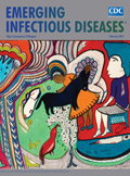
Seeing Things Differently
| EID | Bloom S, Levitt AM. Seeing Things Differently. Emerg Infect Dis. 2014;20(2):340-341. https://doi.org/10.3201/eid2002.ac2002 |
|---|---|
| AMA | Bloom S, Levitt AM. Seeing Things Differently. Emerging Infectious Diseases. 2014;20(2):340-341. doi:10.3201/eid2002.ac2002. |
| APA | Bloom, S., & Levitt, A. M. (2014). Seeing Things Differently. Emerging Infectious Diseases, 20(2), 340-341. https://doi.org/10.3201/eid2002.ac2002. |


