Perspective
Ethics of Infection Control Measures for Carriers of Antimicrobial Drug–Resistant Organisms [PDF - 1.12 MB - 8 pages]
Many countries have implemented infection control measures directed at carriers of multidrug-resistant organisms. To explore the ethical implications of these measures, we analyzed 227 consultations about multidrug resistance and compared them with the literature on communicable disease in general. We found that control measures aimed at carriers have a range of negative implications. Although moral dilemmas seem similar to those encountered while implementing control measures for other infectious diseases, 4 distinct features stand out for carriage of multidrug-resistant organisms: carriage presents itself as a state of being; carriage has limited relevance for the health of the carrier; carriage has little relevance outside healthcare settings; and antimicrobial resistance is a slowly evolving threat on which individual carriers have limited effect. These features are of ethical relevance because they influence the way we traditionally think about infectious disease control and urge us to pay more attention to the personal experience of the individual carrier.
| EID | Rump B, Timen A, Hulscher M, Verweij M. Ethics of Infection Control Measures for Carriers of Antimicrobial Drug–Resistant Organisms. Emerg Infect Dis. 2018;24(9):1609-1616. https://doi.org/10.3201/eid2409.171644 |
|---|---|
| AMA | Rump B, Timen A, Hulscher M, et al. Ethics of Infection Control Measures for Carriers of Antimicrobial Drug–Resistant Organisms. Emerging Infectious Diseases. 2018;24(9):1609-1616. doi:10.3201/eid2409.171644. |
| APA | Rump, B., Timen, A., Hulscher, M., & Verweij, M. (2018). Ethics of Infection Control Measures for Carriers of Antimicrobial Drug–Resistant Organisms. Emerging Infectious Diseases, 24(9), 1609-1616. https://doi.org/10.3201/eid2409.171644. |
Synopses
Travel-Associated Zika Cases and Threat of Local Transmission during Global Outbreak, California, USA [PDF - 1.13 MB - 7 pages]
Zika and associated microcephaly among newborns were reported in Brazil during 2015. Zika has since spread across the Americas, and travel-associated cases were reported throughout the United States. We reviewed travel-associated Zika cases in California to assess the potential threat of local Zika virus transmission, given the regional spread of Aedes aegypti and Ae. albopictus mosquitoes. During November 2015–September 2017, a total of 588 travel-associated Zika cases were reported in California, including 139 infections in pregnant women, 10 congenital infections, and 8 sexually transmitted infections. Most case-patients reported travel to Mexico and Central America, and many returned during a period when they could have been viremic. By September 2017, Ae. aegypti mosquitoes had spread to 124 locations in California, and Ae. albopictus mosquitoes had spread to 53 locations. Continued human and mosquito surveillance and public health education are valuable tools in preventing and detecting Zika virus infections and local transmission in California.
| EID | Porse C, Messenger S, Vugia DJ, Jilek W, Salas M, Watt J, et al. Travel-Associated Zika Cases and Threat of Local Transmission during Global Outbreak, California, USA. Emerg Infect Dis. 2018;24(9):1626-1632. https://doi.org/10.3201/eid2409.180203 |
|---|---|
| AMA | Porse C, Messenger S, Vugia DJ, et al. Travel-Associated Zika Cases and Threat of Local Transmission during Global Outbreak, California, USA. Emerging Infectious Diseases. 2018;24(9):1626-1632. doi:10.3201/eid2409.180203. |
| APA | Porse, C., Messenger, S., Vugia, D. J., Jilek, W., Salas, M., Watt, J....Kramer, V. (2018). Travel-Associated Zika Cases and Threat of Local Transmission during Global Outbreak, California, USA. Emerging Infectious Diseases, 24(9), 1626-1632. https://doi.org/10.3201/eid2409.180203. |
Distinguishing Japanese Spotted Fever and Scrub Typhus, Central Japan, 2004– 2015 [PDF - 1.29 MB - 9 pages]
Japanese spotted fever (JSF) and scrub typhus (ST) are endemic to Japan and share similar clinical features. To document the clinical and epidemiologic characteristics that distinguish these 2 rickettsial diseases, during 2004–2015 we recruited 31 JSF patients, 188 ST patients, and 97 nonrickettsial disease patients from the southern Boso Peninsula of Japan. JSF occurred during April–October and ST during November–December. Patients with JSF and ST were significantly older and more likely to reside in wooded areas than were patients with nonrickettsial diseases. Spatial analyses revealed that JSF and ST clusters rarely overlapped. Clinical findings more frequently observed in JSF than in ST patients were purpura, palmar/plantar rash, hyponatremia, organ damage, and delayed defervescence after treatment. Although their clinical features are similar, JSF and ST differ in seasonality, geographic distribution, physical signs, and severity. Because a considerable percentage of patients did not notice rash and eschar, many rickettsial diseases might be underdiagnosed in Japan.
| EID | Sando E, Suzuki M, Katoh S, Fujita H, Taira M, Yaegashi M, et al. Distinguishing Japanese Spotted Fever and Scrub Typhus, Central Japan, 2004– 2015. Emerg Infect Dis. 2018;24(9):1633-1641. https://doi.org/10.3201/eid2409.171436 |
|---|---|
| AMA | Sando E, Suzuki M, Katoh S, et al. Distinguishing Japanese Spotted Fever and Scrub Typhus, Central Japan, 2004– 2015. Emerging Infectious Diseases. 2018;24(9):1633-1641. doi:10.3201/eid2409.171436. |
| APA | Sando, E., Suzuki, M., Katoh, S., Fujita, H., Taira, M., Yaegashi, M....Ariyoshi, K. (2018). Distinguishing Japanese Spotted Fever and Scrub Typhus, Central Japan, 2004– 2015. Emerging Infectious Diseases, 24(9), 1633-1641. https://doi.org/10.3201/eid2409.171436. |
Systematic Review and Meta-analysis of Postexposure Prophylaxis for Crimean-Congo Hemorrhagic Fever Virus among Healthcare Workers [PDF - 760 KB - 7 pages]
We performed a systematic review and meta-analysis on the effectiveness of ribavirin use for the prevention of infection and death of healthcare workers exposed to patients with Crimean-Congo hemorrhagic fever virus (CCHFV) infection. Splashes with blood or bodily fluids (odds ratio [OR] 4.2), being a nurse or physician (OR 2.1), and treating patients who died from CCHFV infection (OR 3.8) were associated with healthcare workers acquiring CCHFV infection; 7% of the workers who received postexposure prophylaxis (PEP) with ribavirin and 89% of those who did not became infected. PEP with ribavirin reduced the odds of infection (OR 0.01, 95% CI 0–0.03), and ribavirin use <48 hours after symptom onset reduced the odds of death (OR 0.03, 95% CI 0–0.58). The odds of death increased 2.4-fold every day without ribavirin treatment. Ribavirin should be recommended as PEP and early treatment for workers at medium-to-high risk for CCHFV infection.
| EID | Ergönül Ö, Keske Ş, Çeldir M, Kara İ, Pshenichnaya N, Abuova G, et al. Systematic Review and Meta-analysis of Postexposure Prophylaxis for Crimean-Congo Hemorrhagic Fever Virus among Healthcare Workers. Emerg Infect Dis. 2018;24(9):1642-1648. https://doi.org/10.3201/eid2409.171709 |
|---|---|
| AMA | Ergönül Ö, Keske Ş, Çeldir M, et al. Systematic Review and Meta-analysis of Postexposure Prophylaxis for Crimean-Congo Hemorrhagic Fever Virus among Healthcare Workers. Emerging Infectious Diseases. 2018;24(9):1642-1648. doi:10.3201/eid2409.171709. |
| APA | Ergönül, Ö., Keske, Ş., Çeldir, M., Kara, İ., Pshenichnaya, N., Abuova, G....Gönen, M. (2018). Systematic Review and Meta-analysis of Postexposure Prophylaxis for Crimean-Congo Hemorrhagic Fever Virus among Healthcare Workers. Emerging Infectious Diseases, 24(9), 1642-1648. https://doi.org/10.3201/eid2409.171709. |
Event-Based Surveillance at Community and Healthcare Facilities, Vietnam, 2016–2017 [PDF - 1.90 MB - 10 pages]
Surveillance and outbreak reporting systems in Vietnam required improvements to function effectively as early warning and response systems. Accordingly, the Ministry of Health of Vietnam, in collaboration with the US Centers for Disease Control and Prevention, launched a pilot project in 2016 focusing on community and hospital event–based surveillance. The pilot was implemented in 4 of Vietnam’s 63 provinces. The pilot demonstrated that event-based surveillance resulted in early detection and reporting of outbreaks, improved collaboration between the healthcare facilities and preventive sectors of the ministry, and increased community participation in surveillance and reporting.
| EID | Clara A, Do TT, Dao A, Tran PD, Dang TQ, Tran QD, et al. Event-Based Surveillance at Community and Healthcare Facilities, Vietnam, 2016–2017. Emerg Infect Dis. 2018;24(9):1649-1658. https://doi.org/10.3201/eid2409.171851 |
|---|---|
| AMA | Clara A, Do TT, Dao A, et al. Event-Based Surveillance at Community and Healthcare Facilities, Vietnam, 2016–2017. Emerging Infectious Diseases. 2018;24(9):1649-1658. doi:10.3201/eid2409.171851. |
| APA | Clara, A., Do, T. T., Dao, A., Tran, P. D., Dang, T. Q., Tran, Q. D....Balajee, S. (2018). Event-Based Surveillance at Community and Healthcare Facilities, Vietnam, 2016–2017. Emerging Infectious Diseases, 24(9), 1649-1658. https://doi.org/10.3201/eid2409.171851. |
Case Report and Genetic Sequence Analysis of Candidatus Borrelia kalaharica, Southern Africa [PDF - 1.01 MB - 6 pages]
Tickborne relapsing fever caused by Borrelia species is rarely reported in travelers returning from Africa. We report a case of a 71-year-old woman who sought treatment at University Medical Center in Freiburg, Germany, in 2015 with recurrent fever after traveling to southern Africa. We detected spirochetes in Giemsa-stained blood smears. Treatment with doxycycline for suspected tickborne relapsing fever was successful. Sequence analyses of several loci (16S rRNA, flagellin, uvrA) showed high similarity to the recently described Candidatus Borrelia kalaharica, which was found in a traveler returning from the same region earlier that year. We provide additional information regarding the genetic relationship of Candidatus B. kalaharica. Sequence information for an additional 6 housekeeping genes enables improved comparability to other borrelial species that cause relapsing fever. Our report underlines the importance and possible emergence of the only recently delineated pathogen in southern Africa.
| EID | Stete K, Rieg S, Margos G, Häcker G, Wagner D, Kern WV, et al. Case Report and Genetic Sequence Analysis of Candidatus Borrelia kalaharica, Southern Africa. Emerg Infect Dis. 2018;24(9):1659-1664. https://doi.org/10.3201/eid2409.171381 |
|---|---|
| AMA | Stete K, Rieg S, Margos G, et al. Case Report and Genetic Sequence Analysis of Candidatus Borrelia kalaharica, Southern Africa. Emerging Infectious Diseases. 2018;24(9):1659-1664. doi:10.3201/eid2409.171381. |
| APA | Stete, K., Rieg, S., Margos, G., Häcker, G., Wagner, D., Kern, W. V....Fingerle, V. (2018). Case Report and Genetic Sequence Analysis of Candidatus Borrelia kalaharica, Southern Africa. Emerging Infectious Diseases, 24(9), 1659-1664. https://doi.org/10.3201/eid2409.171381. |
We report results from a national surveillance program for Clostridioides difficile infection (CDI) in Sweden, where CDI incidence decreased by 22% and the proportion of multidrug-resistant isolates decreased by 80% during 2012–2016. Variation in incidence between counties also diminished during this period, which might be attributable to implementation of nucleic acid amplification testing as the primary diagnostic tool for most laboratories. In contrast to other studies, our study did not indicate increased CDI incidence attributable the introduction of nucleic acid amplification testing. Our results also suggest that successful implementation of hygiene measures is the major cause of the observed incidence decrease. Despite substantial reductions in CDI incidence and prevalence of multidrug-resistant isolates, Sweden still has one of the highest CDI incidence levels in Europe. This finding is unexpected and warrants further investigation, given that Sweden has among the lowest levels of antimicrobial drug use.
| EID | Rizzardi K, Norén T, Aspevall O, Mäkitalo B, Toepfer M, Johansson Å, et al. National Surveillance for Clostridioides difficile Infection, Sweden, 2009–2016. Emerg Infect Dis. 2018;24(9):1617-1625. https://doi.org/10.3201/eid2409.171658 |
|---|---|
| AMA | Rizzardi K, Norén T, Aspevall O, et al. National Surveillance for Clostridioides difficile Infection, Sweden, 2009–2016. Emerging Infectious Diseases. 2018;24(9):1617-1625. doi:10.3201/eid2409.171658. |
| APA | Rizzardi, K., Norén, T., Aspevall, O., Mäkitalo, B., Toepfer, M., Johansson, Å....Åkerlund, T. (2018). National Surveillance for Clostridioides difficile Infection, Sweden, 2009–2016. Emerging Infectious Diseases, 24(9), 1617-1625. https://doi.org/10.3201/eid2409.171658. |
Research
Novel Orthopoxvirus and Lethal Disease in Cat, Italy [PDF - 3.38 MB - 9 pages]
We report detection and full-genome characterization of a novel orthopoxvirus (OPXV) responsible for a fatal infection in a cat. The virus induced skin lesions histologically characterized by leukocyte infiltration and eosinophilic cytoplasmic inclusions. Different PCR approaches were unable to assign the virus to a defined OPXV species. Large amounts of typical brick-shaped virions, morphologically related to OPXV, were observed by electron microscopy. This OPXV strain (Italy_09/17) was isolated on cell cultures and embryonated eggs. Phylogenetic analysis of 9 concatenated genes showed that this virus was distantly related to cowpox virus, more closely related to to ectromelia virus, and belonged to the same cluster of an OPXV recently isolated from captive macaques in Italy. Extensive epidemiologic surveillance in cats and rodents will assess whether cats are incidental hosts and rodents are the main reservoir of the virus. The zoonotic potential of this novel virus also deserves further investigation.
| EID | Lanave G, Dowgier G, Decaro N, Albanese F, Brogi E, Parisi A, et al. Novel Orthopoxvirus and Lethal Disease in Cat, Italy. Emerg Infect Dis. 2018;24(9):1665-1673. https://doi.org/10.3201/eid2409.171283 |
|---|---|
| AMA | Lanave G, Dowgier G, Decaro N, et al. Novel Orthopoxvirus and Lethal Disease in Cat, Italy. Emerging Infectious Diseases. 2018;24(9):1665-1673. doi:10.3201/eid2409.171283. |
| APA | Lanave, G., Dowgier, G., Decaro, N., Albanese, F., Brogi, E., Parisi, A....Elia, G. (2018). Novel Orthopoxvirus and Lethal Disease in Cat, Italy. Emerging Infectious Diseases, 24(9), 1665-1673. https://doi.org/10.3201/eid2409.171283. |
Emergence of Carbapenemase-Producing Enterobacteriaceae, South-Central Ontario, Canada [PDF - 1.89 MB - 9 pages]
We analyzed population-based surveillance data from the Toronto Invasive Bacterial Diseases Network to describe carbapenemase-producing Enterobacteriaceae (CPE) infections during 2007–2015 in south-central Ontario, Canada. We reviewed patients’ medical records and travel histories, analyzed microbiologic and clinical characteristics of CPE infections, and calculated incidence. Among 291 cases identified, New Delhi metallo-β-lactamase was the predominant carbapenemase (51%). The proportion of CPE-positive patients with prior admission to a hospital in Canada who had not received healthcare abroad or traveled to high-risk areas was 13% for patients with oxacillinase-48, 24% for patients with New Delhi metallo-β-lactamase, 55% for patients with Klebsiella pneumoniae carbapenemase, and 67% for patients with Verona integron-encoded metallo-β-lactamase. Incidence of CPE infection increased, reaching 0.33 cases/100,000 population in 2015. For a substantial proportion of patients, no healthcare abroad or high-risk travel could be established, suggesting CPE acquisition in Canada. Policy and practice changes are needed to mitigate nosocomial CPE transmission in hospitals in Canada.
| EID | Kohler PP, Melano RG, Patel SN, Shafinaz S, Faheem A, Coleman BL, et al. Emergence of Carbapenemase-Producing Enterobacteriaceae, South-Central Ontario, Canada. Emerg Infect Dis. 2018;24(9):1674-1682. https://doi.org/10.3201/eid2409.180164 |
|---|---|
| AMA | Kohler PP, Melano RG, Patel SN, et al. Emergence of Carbapenemase-Producing Enterobacteriaceae, South-Central Ontario, Canada. Emerging Infectious Diseases. 2018;24(9):1674-1682. doi:10.3201/eid2409.180164. |
| APA | Kohler, P. P., Melano, R. G., Patel, S. N., Shafinaz, S., Faheem, A., Coleman, B. L....McGeer, A. (2018). Emergence of Carbapenemase-Producing Enterobacteriaceae, South-Central Ontario, Canada. Emerging Infectious Diseases, 24(9), 1674-1682. https://doi.org/10.3201/eid2409.180164. |
From Culturomics to Clinical Microbiology and Forward [PDF - 670 KB - 8 pages]
Culturomics has permitted discovery of hundreds of new bacterial species isolated from the human microbiome. Profiles generated by using matrix-assisted laser desorption/ionization time-of-flight (MALDI-TOF) mass spectrometry have been added to the mass spectrometer database used in clinical microbiology laboratories. We retrospectively collected raw data from MALDI-TOF mass spectrometry used routinely in our laboratory in Marseille, France, during January 2012–March 2018 and analyzed 16S rDNA sequencing results from misidentified strains. During the study period, 744 species were identified from clinical specimens, of which 21 were species first isolated from culturomics. This collection involved 105 clinical specimens, accounting for 98 patients. In 64 cases, isolation of the bacteria was considered clinically relevant. MALDI-TOF mass spectrometry was able to identify the species in 95.2% of the 105 specimens. While contributing to the extension of the bacterial repertoire associated with humans, culturomics studies also enlarge the spectrum of prokaryotes involved in infectious diseases.
| EID | Dubourg G, Baron S, Cadoret F, Couderc C, Fournier P, Lagier J, et al. From Culturomics to Clinical Microbiology and Forward. Emerg Infect Dis. 2018;24(9):1683-1690. https://doi.org/10.3201/eid2409.170995 |
|---|---|
| AMA | Dubourg G, Baron S, Cadoret F, et al. From Culturomics to Clinical Microbiology and Forward. Emerging Infectious Diseases. 2018;24(9):1683-1690. doi:10.3201/eid2409.170995. |
| APA | Dubourg, G., Baron, S., Cadoret, F., Couderc, C., Fournier, P., Lagier, J....Raoult, D. (2018). From Culturomics to Clinical Microbiology and Forward. Emerging Infectious Diseases, 24(9), 1683-1690. https://doi.org/10.3201/eid2409.170995. |
Dispatches
Association of Batai Virus Infection and Encephalitis in Harbor Seals, Germany, 2016 [PDF - 2.67 MB - 5 pages]
We isolated Batai virus from the brain of a euthanized, 26-year-old, captive harbor seal with meningoencephalomyelitis in Germany. We provide evidence that this orthobunyavirus can naturally infect the central nervous system of a mammal. The full-genome sequence showed differences from a previously reported virus isolate from a mosquito in Germany.
| EID | Jo WK, Pfankuche VM, Lehmbecker A, Martina B, Rubio-Garcia A, Becker S, et al. Association of Batai Virus Infection and Encephalitis in Harbor Seals, Germany, 2016. Emerg Infect Dis. 2018;24(9):1691-1695. https://doi.org/10.3201/eid2409.171829 |
|---|---|
| AMA | Jo WK, Pfankuche VM, Lehmbecker A, et al. Association of Batai Virus Infection and Encephalitis in Harbor Seals, Germany, 2016. Emerging Infectious Diseases. 2018;24(9):1691-1695. doi:10.3201/eid2409.171829. |
| APA | Jo, W. K., Pfankuche, V. M., Lehmbecker, A., Martina, B., Rubio-Garcia, A., Becker, S....Osterhaus, A. (2018). Association of Batai Virus Infection and Encephalitis in Harbor Seals, Germany, 2016. Emerging Infectious Diseases, 24(9), 1691-1695. https://doi.org/10.3201/eid2409.171829. |
Use of Favipiravir to Treat Lassa Virus Infection in Macaques [PDF - 2.14 MB - 4 pages]
Lassa virus, the cause of Lassa fever in humans, is endemic to West Africa. Treatment of Lassa fever is primarily supportive, although ribavirin has shown limited efficacy if administered early during infection. We tested favipiravir in Lassa virus–viremic macaques and found that 300 mg/kg daily for 2 weeks successfully treated infection.
| EID | Rosenke K, Feldmann H, Westover JB, Hanley P, Martellaro C, Feldmann F, et al. Use of Favipiravir to Treat Lassa Virus Infection in Macaques. Emerg Infect Dis. 2018;24(9):1696-1699. https://doi.org/10.3201/eid2409.180233 |
|---|---|
| AMA | Rosenke K, Feldmann H, Westover JB, et al. Use of Favipiravir to Treat Lassa Virus Infection in Macaques. Emerging Infectious Diseases. 2018;24(9):1696-1699. doi:10.3201/eid2409.180233. |
| APA | Rosenke, K., Feldmann, H., Westover, J. B., Hanley, P., Martellaro, C., Feldmann, F....Safronetz, D. (2018). Use of Favipiravir to Treat Lassa Virus Infection in Macaques. Emerging Infectious Diseases, 24(9), 1696-1699. https://doi.org/10.3201/eid2409.180233. |
Aortic Endograft Infection with Mycobacterium chimaera and Granulicatella adiacens, Switzerland, 2014 [PDF - 1.86 MB - 5 pages]
We describe an aortic endograft infection caused by Mycobacterium chimaera and Granulicatella adiacens, successfully treated with prolonged antimicrobial drug therapy after complete explantation of the infected endoprosthesis and extra-anatomical reconstruction. Whole-genome sequencing analysis did not indicate a close relationship to bacterial strains known to cause infections after cardiac surgery.
| EID | Plate A, Kohl TA, Keller PM, Majer S, Fulchini R, Strahm C, et al. Aortic Endograft Infection with Mycobacterium chimaera and Granulicatella adiacens, Switzerland, 2014. Emerg Infect Dis. 2018;24(9):1700-1704. https://doi.org/10.3201/eid2409.180247 |
|---|---|
| AMA | Plate A, Kohl TA, Keller PM, et al. Aortic Endograft Infection with Mycobacterium chimaera and Granulicatella adiacens, Switzerland, 2014. Emerging Infectious Diseases. 2018;24(9):1700-1704. doi:10.3201/eid2409.180247. |
| APA | Plate, A., Kohl, T. A., Keller, P. M., Majer, S., Fulchini, R., Strahm, C....Hasse, B. (2018). Aortic Endograft Infection with Mycobacterium chimaera and Granulicatella adiacens, Switzerland, 2014. Emerging Infectious Diseases, 24(9), 1700-1704. https://doi.org/10.3201/eid2409.180247. |
Estimating Frequency of Probable Autochthonous Cases of Dengue, Japan [PDF - 1.51 MB - 4 pages]
Imported dengue into naive areas is a recognized but unquantified threat. Differentiating imported and autochthonous cases remains problematic. A threshold approach applied to Japan identified several aberrant incidences of dengue. Despite these alerts, no epidemics occurred other than 1 in Yoyogi Park in Tokyo, which was probably an unusual event.
| EID | Senda A, Sakuntabhai A, Inaida S, Teissier Y, Matsuda F, Paul RE. Estimating Frequency of Probable Autochthonous Cases of Dengue, Japan. Emerg Infect Dis. 2018;24(9):1705-1708. https://doi.org/10.3201/eid2409.170408 |
|---|---|
| AMA | Senda A, Sakuntabhai A, Inaida S, et al. Estimating Frequency of Probable Autochthonous Cases of Dengue, Japan. Emerging Infectious Diseases. 2018;24(9):1705-1708. doi:10.3201/eid2409.170408. |
| APA | Senda, A., Sakuntabhai, A., Inaida, S., Teissier, Y., Matsuda, F., & Paul, R. E. (2018). Estimating Frequency of Probable Autochthonous Cases of Dengue, Japan. Emerging Infectious Diseases, 24(9), 1705-1708. https://doi.org/10.3201/eid2409.170408. |
Correlation of Severity of Human Tick-Borne Encephalitis Virus Disease and Pathogenicity in Mice [PDF - 1.08 MB - 4 pages]
We compared 2 tick-borne encephalitis virus strains isolated from 2 different foci that cause different symptoms in tick-borne encephalitis patients, from neurologic to mild gastrointestinal symptoms. We compared neuroinvasiveness, neurovirulence, and proinflammatory cytokine response in mice and found unique differences that contribute to our understanding of pathogenesis.
| EID | Kurhade C, Schreier S, Lee Y, Zegenhagen L, Hjertqvist M, Dobler G, et al. Correlation of Severity of Human Tick-Borne Encephalitis Virus Disease and Pathogenicity in Mice. Emerg Infect Dis. 2018;24(9):1709-1712. https://doi.org/10.3201/eid2409.171825 |
|---|---|
| AMA | Kurhade C, Schreier S, Lee Y, et al. Correlation of Severity of Human Tick-Borne Encephalitis Virus Disease and Pathogenicity in Mice. Emerging Infectious Diseases. 2018;24(9):1709-1712. doi:10.3201/eid2409.171825. |
| APA | Kurhade, C., Schreier, S., Lee, Y., Zegenhagen, L., Hjertqvist, M., Dobler, G....Överby, A. K. (2018). Correlation of Severity of Human Tick-Borne Encephalitis Virus Disease and Pathogenicity in Mice. Emerging Infectious Diseases, 24(9), 1709-1712. https://doi.org/10.3201/eid2409.171825. |
Increasing Prevalence of Borrelia burgdorferi sensu stricto–Infected Blacklegged Ticks in Tennessee Valley, Tennessee, USA [PDF - 2.23 MB - 4 pages]
In 2017, we surveyed forests in the upper Tennessee Valley, Tennessee, USA. We found Ixodes scapularis ticks established in 23 of 26 counties, 4 of which had Borrelia burgdorferi sensu stricto–infected ticks. Public health officials should be vigilant for increasing Lyme disease incidence in this region.
| EID | Hickling GJ, Kelly JR, Auckland LD, Hamer SA. Increasing Prevalence of Borrelia burgdorferi sensu stricto–Infected Blacklegged Ticks in Tennessee Valley, Tennessee, USA. Emerg Infect Dis. 2018;24(9):1713-1716. https://doi.org/10.3201/eid2409.180343 |
|---|---|
| AMA | Hickling GJ, Kelly JR, Auckland LD, et al. Increasing Prevalence of Borrelia burgdorferi sensu stricto–Infected Blacklegged Ticks in Tennessee Valley, Tennessee, USA. Emerging Infectious Diseases. 2018;24(9):1713-1716. doi:10.3201/eid2409.180343. |
| APA | Hickling, G. J., Kelly, J. R., Auckland, L. D., & Hamer, S. A. (2018). Increasing Prevalence of Borrelia burgdorferi sensu stricto–Infected Blacklegged Ticks in Tennessee Valley, Tennessee, USA. Emerging Infectious Diseases, 24(9), 1713-1716. https://doi.org/10.3201/eid2409.180343. |
Susceptibility of White-Tailed Deer to Rift Valley Fever Virus [PDF - 627 KB - 3 pages]
Rift Valley fever virus, a zoonotic arbovirus, poses major health threats to livestock and humans if introduced into the United States. White-tailed deer, which are abundant throughout the country, might be sentinel animals for arboviruses. We determined the susceptibility of these deer to this virus and provide evidence for a potentially major epidemiologic role.
| EID | Wilson WC, Kim I, Trujillo JD, Sunwoo S, Noronha LE, Urbaniak K, et al. Susceptibility of White-Tailed Deer to Rift Valley Fever Virus. Emerg Infect Dis. 2018;24(9):1717-1719. https://doi.org/10.3201/eid2409.180265 |
|---|---|
| AMA | Wilson WC, Kim I, Trujillo JD, et al. Susceptibility of White-Tailed Deer to Rift Valley Fever Virus. Emerging Infectious Diseases. 2018;24(9):1717-1719. doi:10.3201/eid2409.180265. |
| APA | Wilson, W. C., Kim, I., Trujillo, J. D., Sunwoo, S., Noronha, L. E., Urbaniak, K....Richt, J. A. (2018). Susceptibility of White-Tailed Deer to Rift Valley Fever Virus. Emerging Infectious Diseases, 24(9), 1717-1719. https://doi.org/10.3201/eid2409.180265. |
Outbreak of Pneumococcal Meningitis, Paoua Subprefecture, Central African Republic, 2016–2017 [PDF - 533 KB - 3 pages]
We report a pneumococcal meningitis outbreak in the Central African Republic (251 suspected cases; 60 confirmed by latex agglutination test) in 2016–2017. Case-fatality rates (10% for confirmed case-patients) were low. In areas where a recent pneumococcal conjugate vaccine campaign was conducted, a smaller proportion of cases was seen in youngest children.
| EID | Coldiron ME, Touré O, Frank T, Bouygues N, Grais RF. Outbreak of Pneumococcal Meningitis, Paoua Subprefecture, Central African Republic, 2016–2017. Emerg Infect Dis. 2018;24(9):1720-1722. https://doi.org/10.3201/eid2409.171058 |
|---|---|
| AMA | Coldiron ME, Touré O, Frank T, et al. Outbreak of Pneumococcal Meningitis, Paoua Subprefecture, Central African Republic, 2016–2017. Emerging Infectious Diseases. 2018;24(9):1720-1722. doi:10.3201/eid2409.171058. |
| APA | Coldiron, M. E., Touré, O., Frank, T., Bouygues, N., & Grais, R. F. (2018). Outbreak of Pneumococcal Meningitis, Paoua Subprefecture, Central African Republic, 2016–2017. Emerging Infectious Diseases, 24(9), 1720-1722. https://doi.org/10.3201/eid2409.171058. |
Molecular Confirmation of Rocky Mountain Spotted Fever Epidemic Agent in Mexicali, Mexico [PDF - 613 KB - 3 pages]
Since 2008, a large epidemic of Rocky Mountain spotted fever has been emerging among humans and dogs in Mexicali, adjacent to the United States in Baja California, Mexico. We molecularly confirmed the causative agent; this information can be used to study the origin and dynamics of the epidemic.
| EID | Tinoco-Gracia L, Lomelí M, Hori-Oshima S, Stephenson N, Foley J. Molecular Confirmation of Rocky Mountain Spotted Fever Epidemic Agent in Mexicali, Mexico. Emerg Infect Dis. 2018;24(9):1723-1725. https://doi.org/10.3201/eid2409.171523 |
|---|---|
| AMA | Tinoco-Gracia L, Lomelí M, Hori-Oshima S, et al. Molecular Confirmation of Rocky Mountain Spotted Fever Epidemic Agent in Mexicali, Mexico. Emerging Infectious Diseases. 2018;24(9):1723-1725. doi:10.3201/eid2409.171523. |
| APA | Tinoco-Gracia, L., Lomelí, M., Hori-Oshima, S., Stephenson, N., & Foley, J. (2018). Molecular Confirmation of Rocky Mountain Spotted Fever Epidemic Agent in Mexicali, Mexico. Emerging Infectious Diseases, 24(9), 1723-1725. https://doi.org/10.3201/eid2409.171523. |
Fatal Tickborne Phlebovirus Infection in Captive Cheetahs, Japan [PDF - 925 KB - 4 pages]
Two captive cheetahs from a zoo in Japan died of a severe fever with thrombocytopenia syndrome–like illness. Severe fever with thrombocytopenia syndrome virus, an endemic tickborne phlebovirus, was detected systemically with secretion of infectious viruses into the saliva. These cases highlight the risk for exposure of captive animals to endemic arthropodborne pathogens.
| EID | Matsuno K, Nonoue N, Noda A, Kasajima N, Noguchi K, Takano A, et al. Fatal Tickborne Phlebovirus Infection in Captive Cheetahs, Japan. Emerg Infect Dis. 2018;24(9):1726-1729. https://doi.org/10.3201/eid2409.171667 |
|---|---|
| AMA | Matsuno K, Nonoue N, Noda A, et al. Fatal Tickborne Phlebovirus Infection in Captive Cheetahs, Japan. Emerging Infectious Diseases. 2018;24(9):1726-1729. doi:10.3201/eid2409.171667. |
| APA | Matsuno, K., Nonoue, N., Noda, A., Kasajima, N., Noguchi, K., Takano, A....Maeda, K. (2018). Fatal Tickborne Phlebovirus Infection in Captive Cheetahs, Japan. Emerging Infectious Diseases, 24(9), 1726-1729. https://doi.org/10.3201/eid2409.171667. |
Elizabethkingia anophelis and Association with Tap Water and Handwashing, Singapore [PDF - 955 KB - 4 pages]
We report an Elizabethkingia anophelis case cluster associated with contaminated aerators and tap water in a children’s intensive care unit in Singapore in 2017. We demonstrate a likely transmission route for E. anophelis to patients through acquisition of the bacteria on hands of healthcare workers via handwashing.
| EID | Yung C, Maiwald M, Loo LH, Soong HY, Tan CB, Lim PK, et al. Elizabethkingia anophelis and Association with Tap Water and Handwashing, Singapore. Emerg Infect Dis. 2018;24(9):1730-1733. https://doi.org/10.3201/eid2409.171843 |
|---|---|
| AMA | Yung C, Maiwald M, Loo LH, et al. Elizabethkingia anophelis and Association with Tap Water and Handwashing, Singapore. Emerging Infectious Diseases. 2018;24(9):1730-1733. doi:10.3201/eid2409.171843. |
| APA | Yung, C., Maiwald, M., Loo, L. H., Soong, H. Y., Tan, C. B., Lim, P. K....Chan, Y. H. (2018). Elizabethkingia anophelis and Association with Tap Water and Handwashing, Singapore. Emerging Infectious Diseases, 24(9), 1730-1733. https://doi.org/10.3201/eid2409.171843. |
Maripa Virus RNA Load and Antibody Response in Hantavirus Pulmonary Syndrome, French Guiana [PDF - 384 KB - 3 pages]
We report viral RNA loads and antibody responses in 6 severe human cases of Maripa virus infection (2 favorable outcomes) and monitored both measures during the 6-week course of disease in 1 nonfatal case. Further research is needed to determine prevalence of this virus and its effect on other hantaviruses.
| EID | Matheus S, Kallel H, Roux A, Bremand L, Labeau B, Moua D, et al. Maripa Virus RNA Load and Antibody Response in Hantavirus Pulmonary Syndrome, French Guiana. Emerg Infect Dis. 2018;24(9):1734-1736. https://doi.org/10.3201/eid2409.180223 |
|---|---|
| AMA | Matheus S, Kallel H, Roux A, et al. Maripa Virus RNA Load and Antibody Response in Hantavirus Pulmonary Syndrome, French Guiana. Emerging Infectious Diseases. 2018;24(9):1734-1736. doi:10.3201/eid2409.180223. |
| APA | Matheus, S., Kallel, H., Roux, A., Bremand, L., Labeau, B., Moua, D....Lavergne, A. (2018). Maripa Virus RNA Load and Antibody Response in Hantavirus Pulmonary Syndrome, French Guiana. Emerging Infectious Diseases, 24(9), 1734-1736. https://doi.org/10.3201/eid2409.180223. |
Severe Manifestations of Chikungunya Fever in Children, India, 2016 [PDF - 677 KB - 3 pages]
Chikungunya is a relatively benign disease, and a paucity of literature on severe manifestations in children exits. We describe a cohort of pediatric chikungunya fever patients in New Delhi, India, who had severe sepsis and septic shock, which can develop during the acute phase of illness.
| EID | Sharma PK, Kumar M, Aggarwal GK, Kumar V, Srivastava R, Sahani A, et al. Severe Manifestations of Chikungunya Fever in Children, India, 2016. Emerg Infect Dis. 2018;24(9):1737-1739. https://doi.org/10.3201/eid2409.180330 |
|---|---|
| AMA | Sharma PK, Kumar M, Aggarwal GK, et al. Severe Manifestations of Chikungunya Fever in Children, India, 2016. Emerging Infectious Diseases. 2018;24(9):1737-1739. doi:10.3201/eid2409.180330. |
| APA | Sharma, P. K., Kumar, M., Aggarwal, G. K., Kumar, V., Srivastava, R., Sahani, A....Goyal, R. (2018). Severe Manifestations of Chikungunya Fever in Children, India, 2016. Emerging Infectious Diseases, 24(9), 1737-1739. https://doi.org/10.3201/eid2409.180330. |
Zika Virus Seropositivity in 1–4-Year-Old Children, Indonesia, 2014 [PDF - 1.18 MB - 4 pages]
We assessed Zika virus seroprevalence among healthy 1–4-year-old children using a serum sample collection assembled in 2014 representing 30 urban sites across Indonesia. Of 662 samples, 9.1% were Zika virus seropositive, suggesting widespread recent Zika virus transmission and immunity. Larger studies are needed to better determine endemicity in Indonesia.
| EID | Sasmono R, Dhenni R, Yohan B, Pronyk P, Hadinegoro S, Soepardi E, et al. Zika Virus Seropositivity in 1–4-Year-Old Children, Indonesia, 2014. Emerg Infect Dis. 2018;24(9):1740-1743. https://doi.org/10.3201/eid2409.180582 |
|---|---|
| AMA | Sasmono R, Dhenni R, Yohan B, et al. Zika Virus Seropositivity in 1–4-Year-Old Children, Indonesia, 2014. Emerging Infectious Diseases. 2018;24(9):1740-1743. doi:10.3201/eid2409.180582. |
| APA | Sasmono, R., Dhenni, R., Yohan, B., Pronyk, P., Hadinegoro, S., Soepardi, E....Soebandrio, A. (2018). Zika Virus Seropositivity in 1–4-Year-Old Children, Indonesia, 2014. Emerging Infectious Diseases, 24(9), 1740-1743. https://doi.org/10.3201/eid2409.180582. |
Research Letters
Trichodysplasia Spinulosa Polyomavirus in Respiratory Tract of Immunocompromised Child [PDF - 396 KB - 3 pages]
Trichodysplasia spinulosa polyomavirus causes trichodysplasia spinulosa, a skin infection, in immunocompromised persons, but the virus is rarely detected in respiratory samples. Using PCR, we detected persistent virus in respiratory and skin samples from an immunocompromised boy with respiratory signs but no characteristic skin spicules. This virus may play a role in respiratory illness.
| EID | Bagasi AA, Khandaker T, Clark G, Akagha T, Ball JK, Irving WL, et al. Trichodysplasia Spinulosa Polyomavirus in Respiratory Tract of Immunocompromised Child. Emerg Infect Dis. 2018;24(9):1744-1746. https://doi.org/10.3201/eid2409.180829 |
|---|---|
| AMA | Bagasi AA, Khandaker T, Clark G, et al. Trichodysplasia Spinulosa Polyomavirus in Respiratory Tract of Immunocompromised Child. Emerging Infectious Diseases. 2018;24(9):1744-1746. doi:10.3201/eid2409.180829. |
| APA | Bagasi, A. A., Khandaker, T., Clark, G., Akagha, T., Ball, J. K., Irving, W. L....McClure, C. (2018). Trichodysplasia Spinulosa Polyomavirus in Respiratory Tract of Immunocompromised Child. Emerging Infectious Diseases, 24(9), 1744-1746. https://doi.org/10.3201/eid2409.180829. |
Wohlfahrtiimonas chitiniclastica Bacteremia in Hospitalized Homeless Man with Squamous Cell Carcinoma [PDF - 369 KB - 3 pages]
We report a case of Wohlfahrtiimonas chitiniclastica bacteremia in an elderly man in Japan who had squamous cell carcinoma. Blood cultures were initially negative for W. chitiniclastica but were positive on day 20. Careful attention needs to be paid to this organism in patients who have chronic wounds with maggots.
| EID | Katanami Y, Kutsuna S, Nagashima M, Takaya S, Yamamoto K, Takeshita N, et al. Wohlfahrtiimonas chitiniclastica Bacteremia in Hospitalized Homeless Man with Squamous Cell Carcinoma. Emerg Infect Dis. 2018;24(9):1746-1748. https://doi.org/10.3201/eid2409.170080 |
|---|---|
| AMA | Katanami Y, Kutsuna S, Nagashima M, et al. Wohlfahrtiimonas chitiniclastica Bacteremia in Hospitalized Homeless Man with Squamous Cell Carcinoma. Emerging Infectious Diseases. 2018;24(9):1746-1748. doi:10.3201/eid2409.170080. |
| APA | Katanami, Y., Kutsuna, S., Nagashima, M., Takaya, S., Yamamoto, K., Takeshita, N....Ohmagari, N. (2018). Wohlfahrtiimonas chitiniclastica Bacteremia in Hospitalized Homeless Man with Squamous Cell Carcinoma. Emerging Infectious Diseases, 24(9), 1746-1748. https://doi.org/10.3201/eid2409.170080. |
Symptomatic Dengue during Pregnancy and Congenital Neurologic Malformations [PDF - 316 KB - 3 pages]
Dengue virus infection during pregnancy increased the risk for any neurologic congenital anomaly in the infant by roughly 50% and for other congenital malformations of brain 4-fold. Our results show an association between dengue during pregnancy and congenital anomalies of the brain, suggesting that flaviviruses other than Zika virus are associated with such malformations.
| EID | Paixão ES, Teixeira M, Costa MN, Barreto ML, Rodrigues LC. Symptomatic Dengue during Pregnancy and Congenital Neurologic Malformations. Emerg Infect Dis. 2018;24(9):1748-1750. https://doi.org/10.3201/eid2409.170361 |
|---|---|
| AMA | Paixão ES, Teixeira M, Costa MN, et al. Symptomatic Dengue during Pregnancy and Congenital Neurologic Malformations. Emerging Infectious Diseases. 2018;24(9):1748-1750. doi:10.3201/eid2409.170361. |
| APA | Paixão, E. S., Teixeira, M., Costa, M. N., Barreto, M. L., & Rodrigues, L. C. (2018). Symptomatic Dengue during Pregnancy and Congenital Neurologic Malformations. Emerging Infectious Diseases, 24(9), 1748-1750. https://doi.org/10.3201/eid2409.170361. |
Travelers’ Actual and Subjective Knowledge about Risk for Ebola Virus Disease [PDF - 340 KB - 2 pages]
To determine travelers’ actual and subjective knowledge about risk for Ebola virus disease, we surveyed travelers from France. Actual knowledge did not prevent irrational perceptions or promote safe behavior. Rather, readiness to adopt protective behavior depended on subjective knowledge and overconfidence in ability to self-protect.
| EID | Régner I, Ianos O, Shajrawi L, Brouqui P, Gautret P. Travelers’ Actual and Subjective Knowledge about Risk for Ebola Virus Disease. Emerg Infect Dis. 2018;24(9):1750-1751. https://doi.org/10.3201/eid2409.171343 |
|---|---|
| AMA | Régner I, Ianos O, Shajrawi L, et al. Travelers’ Actual and Subjective Knowledge about Risk for Ebola Virus Disease. Emerging Infectious Diseases. 2018;24(9):1750-1751. doi:10.3201/eid2409.171343. |
| APA | Régner, I., Ianos, O., Shajrawi, L., Brouqui, P., & Gautret, P. (2018). Travelers’ Actual and Subjective Knowledge about Risk for Ebola Virus Disease. Emerging Infectious Diseases, 24(9), 1750-1751. https://doi.org/10.3201/eid2409.171343. |
Spread of mcr-1–Driven Colistin Resistance on Hospital Surfaces, Italy [PDF - 395 KB - 2 pages]
Plasmid-mediated colistin resistance driven by the mcr-1 gene is of great clinical concern. Its diffusion in the hospital environment is unknown. We detected mcr-1–driven resistance in 8.3% of Enterobacteriaceae isolates from hospital surfaces in Italy, which might represent a reservoir of threatening nosocomial pathogens.
| EID | Caselli E, D’Accolti M, Soffritti I, Piffanelli M, Mazzacane S. Spread of mcr-1–Driven Colistin Resistance on Hospital Surfaces, Italy. Emerg Infect Dis. 2018;24(9):1752-1753. https://doi.org/10.3201/eid2409.171386 |
|---|---|
| AMA | Caselli E, D’Accolti M, Soffritti I, et al. Spread of mcr-1–Driven Colistin Resistance on Hospital Surfaces, Italy. Emerging Infectious Diseases. 2018;24(9):1752-1753. doi:10.3201/eid2409.171386. |
| APA | Caselli, E., D’Accolti, M., Soffritti, I., Piffanelli, M., & Mazzacane, S. (2018). Spread of mcr-1–Driven Colistin Resistance on Hospital Surfaces, Italy. Emerging Infectious Diseases, 24(9), 1752-1753. https://doi.org/10.3201/eid2409.171386. |
Transverse Myelitis and Guillain-Barré Syndrome Associated with Cat-Scratch Disease, Texas, USA, 2011 [PDF - 404 KB - 2 pages]
We describe a case of coexisting transverse myelitis and Guillain-Barré syndrome related to infection with Bartonella henselae proteobacterium and review similar serology-proven cases. B. henselae infection might be emerging as a cause of myelitis and Guillain-Barré syndrome and should be considered as an etiologic factor in patients with such clinical presentations.
| EID | Zakhour R, Mancias P, Heresi G, Pérez N. Transverse Myelitis and Guillain-Barré Syndrome Associated with Cat-Scratch Disease, Texas, USA, 2011. Emerg Infect Dis. 2018;24(9):1754-1755. https://doi.org/10.3201/eid2409.180008 |
|---|---|
| AMA | Zakhour R, Mancias P, Heresi G, et al. Transverse Myelitis and Guillain-Barré Syndrome Associated with Cat-Scratch Disease, Texas, USA, 2011. Emerging Infectious Diseases. 2018;24(9):1754-1755. doi:10.3201/eid2409.180008. |
| APA | Zakhour, R., Mancias, P., Heresi, G., & Pérez, N. (2018). Transverse Myelitis and Guillain-Barré Syndrome Associated with Cat-Scratch Disease, Texas, USA, 2011. Emerging Infectious Diseases, 24(9), 1754-1755. https://doi.org/10.3201/eid2409.180008. |
Co-Circulation of 4 Dengue Virus Serotypes among Travelers Entering China from Myanmar, 2017 [PDF - 634 KB - 3 pages]
We report 301 dengue virus infections among cross-border travelers entering Yunnan Province, China, from Myanmar during 2017. Phylogenetic analysis of 99 strains found all 4 serotypes co-circulating; genetic characteristics have also changed. This finding highlights the urgent need for monitoring dengue virus cross-border transmission as early warning of severe dengue fever.
| EID | Wang B, Liang Y, Yang S, Du Y, Xiong L, Zhao T, et al. Co-Circulation of 4 Dengue Virus Serotypes among Travelers Entering China from Myanmar, 2017. Emerg Infect Dis. 2018;24(9):1756-1758. https://doi.org/10.3201/eid2409.180252 |
|---|---|
| AMA | Wang B, Liang Y, Yang S, et al. Co-Circulation of 4 Dengue Virus Serotypes among Travelers Entering China from Myanmar, 2017. Emerging Infectious Diseases. 2018;24(9):1756-1758. doi:10.3201/eid2409.180252. |
| APA | Wang, B., Liang, Y., Yang, S., Du, Y., Xiong, L., Zhao, T....Xia, X. (2018). Co-Circulation of 4 Dengue Virus Serotypes among Travelers Entering China from Myanmar, 2017. Emerging Infectious Diseases, 24(9), 1756-1758. https://doi.org/10.3201/eid2409.180252. |
Case of Microcephaly after Congenital Infection with Asian Lineage Zika Virus, Thailand [PDF - 1.42 MB - 4 pages]
We sequenced the virus genomes from 3 pregnant women in Thailand with Zika virus diagnoses. All had infections with the Asian lineage. The woman infected at gestational week 9, and not those infected at weeks 20 and 24, had a fetus with microcephaly. Asian lineage Zika viruses can cause microcephaly.
| EID | Wongsurawat T, Athipanyasilp N, Jenjaroenpun P, Jun S, Kaewnapan B, Wassenaar TM, et al. Case of Microcephaly after Congenital Infection with Asian Lineage Zika Virus, Thailand. Emerg Infect Dis. 2018;24(9):1758-1761. https://doi.org/10.3201/eid2409.180416 |
|---|---|
| AMA | Wongsurawat T, Athipanyasilp N, Jenjaroenpun P, et al. Case of Microcephaly after Congenital Infection with Asian Lineage Zika Virus, Thailand. Emerging Infectious Diseases. 2018;24(9):1758-1761. doi:10.3201/eid2409.180416. |
| APA | Wongsurawat, T., Athipanyasilp, N., Jenjaroenpun, P., Jun, S., Kaewnapan, B., Wassenaar, T. M....Horthongkham, N. (2018). Case of Microcephaly after Congenital Infection with Asian Lineage Zika Virus, Thailand. Emerging Infectious Diseases, 24(9), 1758-1761. https://doi.org/10.3201/eid2409.180416. |
Dirofilaria repens Nematode Infection with Microfilaremia in Traveler Returning to Belgium from Senegal [PDF - 736 KB - 3 pages]
We report human infection with a Dirofilaria repens nematode likely acquired in Senegal. An adult worm was extracted from the right conjunctiva of the case-patient, and blood microfilariae were detected, which led to an initial misdiagnosis of loiasis. We also observed the complete life cycle of a D. repens nematode in this patient.
| EID | Potters I, Vanfraechem G, Bottieau E. Dirofilaria repens Nematode Infection with Microfilaremia in Traveler Returning to Belgium from Senegal. Emerg Infect Dis. 2018;24(9):1761-1763. https://doi.org/10.3201/eid2409.180462 |
|---|---|
| AMA | Potters I, Vanfraechem G, Bottieau E. Dirofilaria repens Nematode Infection with Microfilaremia in Traveler Returning to Belgium from Senegal. Emerging Infectious Diseases. 2018;24(9):1761-1763. doi:10.3201/eid2409.180462. |
| APA | Potters, I., Vanfraechem, G., & Bottieau, E. (2018). Dirofilaria repens Nematode Infection with Microfilaremia in Traveler Returning to Belgium from Senegal. Emerging Infectious Diseases, 24(9), 1761-1763. https://doi.org/10.3201/eid2409.180462. |
Rubella Virus Genotype 1E in Travelers Returning to Japan from Indonesia, 2017 [PDF - 573 KB - 3 pages]
Although rubella is epidemic in Indonesia, the phylogenetic profile of circulating rubella virus strains has not been clarified. In 2017, rubella virus was detected in 2 travelers who returned from Indonesia to Japan. These strains were classified into genotype 1E lineage 2, which may be an indigenous strain in Indonesia.
| EID | Kanbayashi D, Kurata T, Nishino Y, Orii F, Takii Y, Kinoshita M, et al. Rubella Virus Genotype 1E in Travelers Returning to Japan from Indonesia, 2017. Emerg Infect Dis. 2018;24(9):1763-1765. https://doi.org/10.3201/eid2409.180621 |
|---|---|
| AMA | Kanbayashi D, Kurata T, Nishino Y, et al. Rubella Virus Genotype 1E in Travelers Returning to Japan from Indonesia, 2017. Emerging Infectious Diseases. 2018;24(9):1763-1765. doi:10.3201/eid2409.180621. |
| APA | Kanbayashi, D., Kurata, T., Nishino, Y., Orii, F., Takii, Y., Kinoshita, M....Yumisashi, T. (2018). Rubella Virus Genotype 1E in Travelers Returning to Japan from Indonesia, 2017. Emerging Infectious Diseases, 24(9), 1763-1765. https://doi.org/10.3201/eid2409.180621. |
Spondweni Virus in Field-Caught Culex quinquefasciatus Mosquitoes, Haiti, 2016 [PDF - 349 KB - 3 pages]
Spondweni virus (SPONV) and Zika virus cause similar diseases in humans. We detected SPONV outside of Africa from a pool of Culex mosquitoes collected in Haiti in 2016. This finding raises questions about the role of SPONV as a human pathogen in Haiti and other Caribbean countries.
| EID | White SK, Lednicky JA, Okech BA, Morris J, Dunford JC. Spondweni Virus in Field-Caught Culex quinquefasciatus Mosquitoes, Haiti, 2016. Emerg Infect Dis. 2018;24(9):1765-1767. https://doi.org/10.3201/eid2409.171957 |
|---|---|
| AMA | White SK, Lednicky JA, Okech BA, et al. Spondweni Virus in Field-Caught Culex quinquefasciatus Mosquitoes, Haiti, 2016. Emerging Infectious Diseases. 2018;24(9):1765-1767. doi:10.3201/eid2409.171957. |
| APA | White, S. K., Lednicky, J. A., Okech, B. A., Morris, J., & Dunford, J. C. (2018). Spondweni Virus in Field-Caught Culex quinquefasciatus Mosquitoes, Haiti, 2016. Emerging Infectious Diseases, 24(9), 1765-1767. https://doi.org/10.3201/eid2409.171957. |
Fluconazole-Resistant Candida parapsilosis Bloodstream Isolates with Y132F Mutation in ERG11 Gene, South Korea [PDF - 395 KB - 3 pages]
We recently observed the emergence of fluconazole-resistant Candida parapsilosis bloodstream isolates harboring a Y132F substitution in Erg11p in South Korea. These Y132F isolates had a higher propensity to cause clonal transmission than other fluconazole-resistant isolates and persisted within hospitals for several years, as revealed by microsatellite typing.
| EID | Choi Y, Kim Y, Yong D, Byun J, Kim T, Chang Y, et al. Fluconazole-Resistant Candida parapsilosis Bloodstream Isolates with Y132F Mutation in ERG11 Gene, South Korea. Emerg Infect Dis. 2018;24(9):1768-1770. https://doi.org/10.3201/eid2409.180625 |
|---|---|
| AMA | Choi Y, Kim Y, Yong D, et al. Fluconazole-Resistant Candida parapsilosis Bloodstream Isolates with Y132F Mutation in ERG11 Gene, South Korea. Emerging Infectious Diseases. 2018;24(9):1768-1770. doi:10.3201/eid2409.180625. |
| APA | Choi, Y., Kim, Y., Yong, D., Byun, J., Kim, T., Chang, Y....Shin, J. (2018). Fluconazole-Resistant Candida parapsilosis Bloodstream Isolates with Y132F Mutation in ERG11 Gene, South Korea. Emerging Infectious Diseases, 24(9), 1768-1770. https://doi.org/10.3201/eid2409.180625. |
Borrelia miyamotoi Disease in an Immunocompetent Patient, Western Europe [PDF - 514 KB - 3 pages]
Borrelia miyamotoi disease is a hard tick–borne relapsing fever illness that occurs across the temperate climate zone. Human B. miyamotoi disease in immunocompetent patients has been described in Russia, North America, and Japan. We describe a case of B. miyamotoi disease in an immunocompetent patient in western Europe.
| EID | Hoornstra D, Koetsveld J, Sprong H, Platonov AE, Hovius JW. Borrelia miyamotoi Disease in an Immunocompetent Patient, Western Europe. Emerg Infect Dis. 2018;24(9):1770-1772. https://doi.org/10.3201/eid2409.180806 |
|---|---|
| AMA | Hoornstra D, Koetsveld J, Sprong H, et al. Borrelia miyamotoi Disease in an Immunocompetent Patient, Western Europe. Emerging Infectious Diseases. 2018;24(9):1770-1772. doi:10.3201/eid2409.180806. |
| APA | Hoornstra, D., Koetsveld, J., Sprong, H., Platonov, A. E., & Hovius, J. W. (2018). Borrelia miyamotoi Disease in an Immunocompetent Patient, Western Europe. Emerging Infectious Diseases, 24(9), 1770-1772. https://doi.org/10.3201/eid2409.180806. |
Letters
Seroprevalence of Chikungunya Virus after Its Emergence in Brazil [PDF - 305 KB - 1 page]
| EID | Gérardin P, Freitas A, Sissoko D. Seroprevalence of Chikungunya Virus after Its Emergence in Brazil. Emerg Infect Dis. 2018;24(9):1773. https://doi.org/10.3201/eid2409.180724 |
|---|---|
| AMA | Gérardin P, Freitas A, Sissoko D. Seroprevalence of Chikungunya Virus after Its Emergence in Brazil. Emerging Infectious Diseases. 2018;24(9):1773. doi:10.3201/eid2409.180724. |
| APA | Gérardin, P., Freitas, A., & Sissoko, D. (2018). Seroprevalence of Chikungunya Virus after Its Emergence in Brazil. Emerging Infectious Diseases, 24(9), 1773. https://doi.org/10.3201/eid2409.180724. |
Another Dimension
Emerging Infectious Literatures and the Zombie Condition [PDF - 370 KB - 5 pages]
The book club format has enabled expert and nonexpert exploration of infection and epidemiology as encountered in popular literature. This exploration reveals that fiction focusing on apocalyptic disease often uses the zombie as embodiment of infection, as well as an exemplar of current knowledge on emerging disease.
| EID | Verran J, Reyes X. Emerging Infectious Literatures and the Zombie Condition. Emerg Infect Dis. 2018;24(9):1774-1778. https://doi.org/10.3201/eid2409.170658 |
|---|---|
| AMA | Verran J, Reyes X. Emerging Infectious Literatures and the Zombie Condition. Emerging Infectious Diseases. 2018;24(9):1774-1778. doi:10.3201/eid2409.170658. |
| APA | Verran, J., & Reyes, X. (2018). Emerging Infectious Literatures and the Zombie Condition. Emerging Infectious Diseases, 24(9), 1774-1778. https://doi.org/10.3201/eid2409.170658. |
Etymologia
Etymologia: Granulicatella [PDF - 485 KB - 1 page]
| EID | Henry R. Etymologia: Granulicatella. Emerg Infect Dis. 2018;24(9):1704. https://doi.org/10.3201/eid2409.et2409 |
|---|---|
| AMA | Henry R. Etymologia: Granulicatella. Emerging Infectious Diseases. 2018;24(9):1704. doi:10.3201/eid2409.et2409. |
| APA | Henry, R. (2018). Etymologia: Granulicatella. Emerging Infectious Diseases, 24(9), 1704. https://doi.org/10.3201/eid2409.et2409. |
Online Reports
World Health Organization Methodology to Prioritize Emerging Infectious Diseases in Need of Research and Development [PDF - 1.04 MB - 10 pages]
The World Health Organization R&D Blueprint aims to accelerate the availability of medical technologies during epidemics by focusing on a list of prioritized emerging diseases for which medical countermeasures are insufficient or nonexistent. The prioritization process has 3 components: a Delphi process to narrow down a list of potential priority diseases, a multicriteria decision analysis to rank the short list of diseases, and a final Delphi round to arrive at a final list of 10 diseases. A group of international experts applied this process in January 2017, resulting in a list of 10 priority diseases. The robustness of the list was tested by performing a sensitivity analysis. The new process corrected major shortcomings in the pre–R&D Blueprint approach to disease prioritization and increased confidence in the results.
Corrections
Correction: Vol. 24, No. 4 [PDF - 138 KB - 1 page]
Correction: Vol. 24, No. 8 [PDF - 138 KB - 1 page]
About the Cover
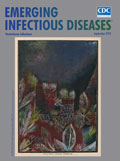
Tropical Sunset Blues [PDF - 2.70 MB - 2 pages]
| EID | Breedlove B, Arguin PM. Tropical Sunset Blues. Emerg Infect Dis. 2018;24(9):1779-1780. https://doi.org/10.3201/eid2409.ac2409 |
|---|---|
| AMA | Breedlove B, Arguin PM. Tropical Sunset Blues. Emerging Infectious Diseases. 2018;24(9):1779-1780. doi:10.3201/eid2409.ac2409. |
| APA | Breedlove, B., & Arguin, P. M. (2018). Tropical Sunset Blues. Emerging Infectious Diseases, 24(9), 1779-1780. https://doi.org/10.3201/eid2409.ac2409. |
News and Notes
International Conference on Emerging Infectious Diseases 2018 Poster and Oral Presentation Abstracts [PDF - 2.96 MB - 247 pages]
| EID | International Conference on Emerging Infectious Diseases 2018 Poster and Oral Presentation Abstracts. Emerg Infect Dis. 2018;24(9):1. https://doi.org/10.3201/eid2409.nn2409 |
|---|---|
| AMA | International Conference on Emerging Infectious Diseases 2018 Poster and Oral Presentation Abstracts. Emerging Infectious Diseases. 2018;24(9):1. doi:10.3201/eid2409.nn2409. |
| APA | (2018). International Conference on Emerging Infectious Diseases 2018 Poster and Oral Presentation Abstracts. Emerging Infectious Diseases, 24(9), 1. https://doi.org/10.3201/eid2409.nn2409. |


