Volume 10, Number 11—November 2004
Research
Topographic Changes in SARS Coronavirus–infected Cells during Late Stages of Infection
Mah-Lee Ng* , J.W.M. Lee*, M.L.N. Leong*, A.-E. Ling†, H.-C. Tan‡, and E.E. Ooi‡
Figure 4
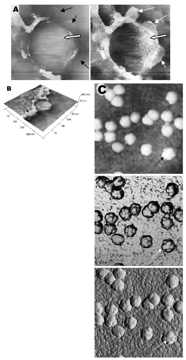
Figure 4. Atomic force microscopy of Vero cells infected with severe acute respiratory syndrome–associated coronavirus at 15 h after infection. A) At much higher resolution imaging of the edge of a cell, a virus particle (thick arrow) in the process of extruding from the cell plasma membrane (PM) after fusion of the transport vesicle with the cell membrane. PM shows some loss of integrity (thin arrows) during this exit process. B) A three-dimensional reconstruction of the extruding virus particle from panel B. Arrow indicates the thickened cell edge.
Page created: April 21, 2011
Page updated: April 21, 2011
Page reviewed: April 21, 2011
The conclusions, findings, and opinions expressed by authors contributing to this journal do not necessarily reflect the official position of the U.S. Department of Health and Human Services, the Public Health Service, the Centers for Disease Control and Prevention, or the authors' affiliated institutions. Use of trade names is for identification only and does not imply endorsement by any of the groups named above.