Volume 29, Number 1—January 2023
Dispatch
Photobacterium damselae subspecies damselae Pneumonia in Dead, Stranded Bottlenose Dolphin, Eastern Mediterranean Sea
Danny Morick , Shlomo E. Blum, Nadav Davidovich, Ziv Zemah-Shamir, Eyal Bigal, Peleg Itay, Assaf Rokney, Iris Nasie, Noa Feldman, Marcelo Flecker, Mia Roditi-Elasar, Kobi Aharoni, Yotam Zuriel, Natascha Wosnick, Dan Tchernov, and Aviad P. Scheinin
Figure 1
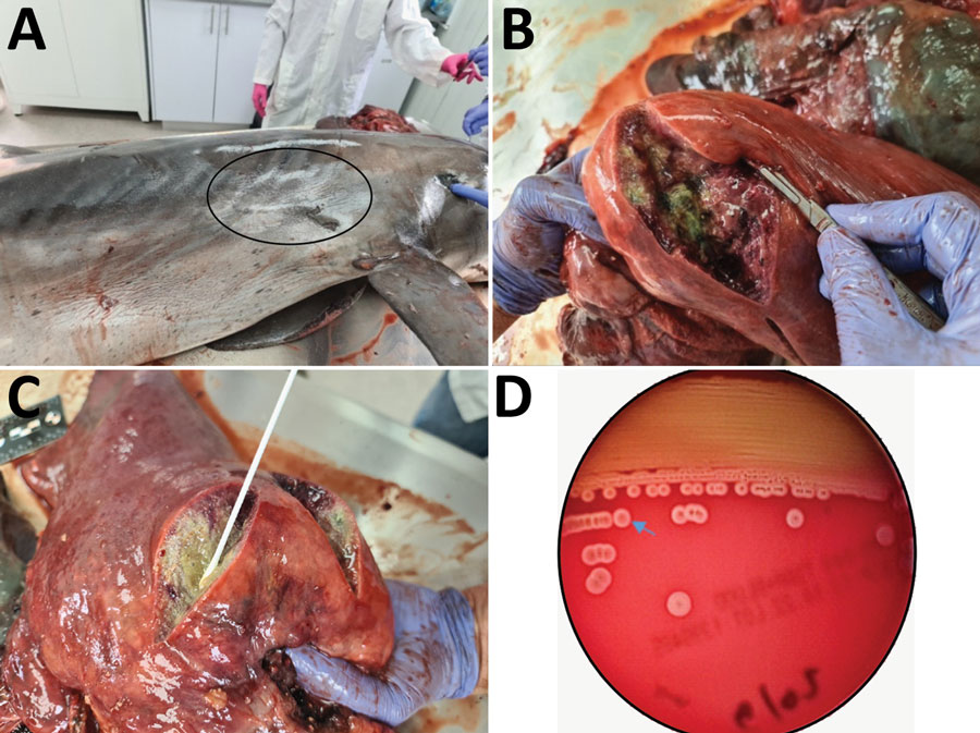
Figure 1. Photobacterium damselae subspecies damselae pneumonia in a bottlenose dolphin, eastern Mediterranean Sea. Gross pathologic examination of the dolphin (Tursiops truncatus) showed a scar (oval) at the right side of the chest (A) that might be a sign for a previous wound that initiated the infection (B, C). Four abscesses, 5–10 cm in diameter, filled with purulent fluid and necrotic debris were observed in the right lung of the animal. Hemolytic phenotype of the P. damselae subsp. damselae isolate on sheep blood agar (D) indicates the border of the halo of 1 colony (arrow). A weak hemolytic phenotype was observed after culturing isolate on blood agar plates for 24 h.
Page created: October 20, 2022
Page updated: December 22, 2022
Page reviewed: December 22, 2022
The conclusions, findings, and opinions expressed by authors contributing to this journal do not necessarily reflect the official position of the U.S. Department of Health and Human Services, the Public Health Service, the Centers for Disease Control and Prevention, or the authors' affiliated institutions. Use of trade names is for identification only and does not imply endorsement by any of the groups named above.