Photobacterium damselae subspecies damselae Pneumonia in Dead, Stranded Bottlenose Dolphin, Eastern Mediterranean Sea
Danny Morick

, Shlomo E. Blum, Nadav Davidovich, Ziv Zemah-Shamir, Eyal Bigal, Peleg Itay, Assaf Rokney, Iris Nasie, Noa Feldman, Marcelo Flecker, Mia Roditi-Elasar, Kobi Aharoni, Yotam Zuriel, Natascha Wosnick, Dan Tchernov, and Aviad P. Scheinin
Author affiliations: University of Haifa, Haifa, Israel (D. Morick, N. Davidovich, Z. Zemah-Shamir, E. Bigal, P. Itay, M. Roditi-Elasar, Y. Zuriel, D. Tchernov, A.P. Scheinin); Hong Kong Branch of Southern Marine Science and Engineering, Guangzhou, China (D. Morick, D. Tchernov); Kimron Veterinary Institute, Bet Dagan, Israel (S.E. Blum, M. Flecker); Israeli Veterinary Services, Bet Dagan (N. Davidovich); Ministry of Health, Jerusalem, Israel (A. Rokney, I. Nasie, N. Feldman); Hebrew University of Jerusalem, Rehovot, Israel (K. Aharoni); Universidade Federal do Paraná, Curitiba, Brazil (N. Wosnick)
Main Article
Figure 2
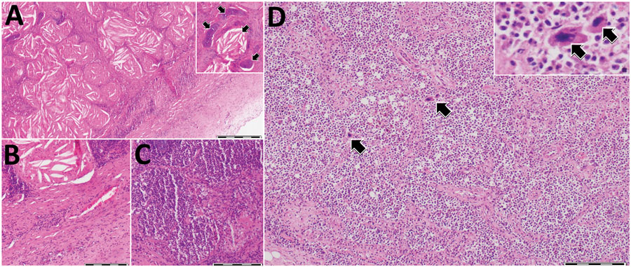
Figure 2. Histologic analysis of lungs and spleen of a bottlenose dolphin (Tursiops truncatus) with Photobacterium damselae subspecies damselae pneumonia, eastern Mediterranean Sea. A) Lung tissue showed a nodular structure covered by fibrous capsule (right bottom of figure panel) composed of numerous cholesterol clefts and areas of reactive fibrosis. Hyaline cartilage was observed, interpreted as bronchi and bronchioles. Inset, higher magnification showing an aggregate of cholesterol clefts and hyaline cartilage (arrow). B) Abundant fibrous lung tissue (lower right half) and cellular infiltrates were also observed. C) Different area of the lung parenchyma characterized by increased cellular infiltration. D) Spleen expressed an apparent contraction of the parenchyma showing diffuse cellularity, with only a few defined lymphoid follicles, as well as megakaryocytes (arrows) indicative of extramedullary hematopoiesis. Inset: higher magnification showing 2 adjacent megakaryocytes (arrows). Hematoxylin and eosin stained. Scale bars indicate 500 μm in panel A and 200 μm in panels B–D.
Main Article
Page created: October 20, 2022
Page updated: December 22, 2022
Page reviewed: December 22, 2022
The conclusions, findings, and opinions expressed by authors contributing to this journal do not necessarily reflect the official position of the U.S. Department of Health and Human Services, the Public Health Service, the Centers for Disease Control and Prevention, or the authors' affiliated institutions. Use of trade names is for identification only and does not imply endorsement by any of the groups named above.