Perspective
Leveraging Advances in Tuberculosis Diagnosis and Treatment to Address Nontuberculous Mycobacterial Disease [PDF - 500 KB - 5 pages]
The nontuberculous mycobacteria (NTM), defined as any mycobacterial pathogen other than Mycobacterium tuberculosis or Mycobacterium leprae, are a diverse group of pathogens that collectively cause a substantive but often unappreciated worldwide burden of illness. Although NTMs may cause illness similar to M. tuberculosis, these pathogens generally do not respond to classic tuberculosis (TB) drug regimens, resulting in misdiagnosis and poor treatment, particularly in resource-poor settings. Although a few high-quality epidemiologic surveys have been made on the topic, existing evidence suggests that NTM-associated disease is much more common than previously thought: more common than TB in the industrialized world and likely increasing in prevalence globally. Despite this evidence, these organisms remain markedly understudied, and few international grants support basic science and clinical research. Here we suggest that the considerable efforts in developing new treatments and diagnostics for TB can be harnessed in the fight against NTM-associated illnesses.
| EID | Raju RM, Raju SM, Zhao Y, Rubin EJ. Leveraging Advances in Tuberculosis Diagnosis and Treatment to Address Nontuberculous Mycobacterial Disease. Emerg Infect Dis. 2016;22(3):365-369. https://doi.org/10.3201/eid2203.151643 |
|---|---|
| AMA | Raju RM, Raju SM, Zhao Y, et al. Leveraging Advances in Tuberculosis Diagnosis and Treatment to Address Nontuberculous Mycobacterial Disease. Emerging Infectious Diseases. 2016;22(3):365-369. doi:10.3201/eid2203.151643. |
| APA | Raju, R. M., Raju, S. M., Zhao, Y., & Rubin, E. J. (2016). Leveraging Advances in Tuberculosis Diagnosis and Treatment to Address Nontuberculous Mycobacterial Disease. Emerging Infectious Diseases, 22(3), 365-369. https://doi.org/10.3201/eid2203.151643. |
Synopses
Histoplasmosis has been described as the most common endemic mycosis in the United States. However, histoplasmosis is not nationally notifiable. Its presumed geographic distribution is largely derived from skin test surveys performed during the 1940s, and information about its local features comes primarily from outbreak investigations. We conducted a literature review to assess epidemiologic features of histoplasmosis outbreaks in the United States. During 1938–2013, a total of 105 outbreaks involving 2,850 cases were reported in 26 states and the territory of Puerto Rico. Common exposure settings were chicken coops and buildings or other structures undergoing renovation or demolition. Birds, bats, or their droppings were reported to be present in 77% of outbreak settings, and workplace exposures were reported in 41% of outbreaks. The continued occurrence of histoplasmosis outbreaks, particularly work-related ones involving known disturbance of bird or bat droppings, highlights the need to increase awareness of the disease.
| EID | Benedict K, Mody RK. Epidemiology of Histoplasmosis Outbreaks, United States, 1938–2013. Emerg Infect Dis. 2016;22(3):370-378. https://doi.org/10.3201/eid2203.151117 |
|---|---|
| AMA | Benedict K, Mody RK. Epidemiology of Histoplasmosis Outbreaks, United States, 1938–2013. Emerging Infectious Diseases. 2016;22(3):370-378. doi:10.3201/eid2203.151117. |
| APA | Benedict, K., & Mody, R. K. (2016). Epidemiology of Histoplasmosis Outbreaks, United States, 1938–2013. Emerging Infectious Diseases, 22(3), 370-378. https://doi.org/10.3201/eid2203.151117. |
Avian Influenza A(H5N1) Virus in Egypt [PDF - 854 KB - 10 pages]
In Egypt, avian influenza A subtype H5N1 and H9N2 viruses are enzootic in poultry. The control plan devised by veterinary authorities in Egypt to prevent infections in poultry focused mainly on vaccination and ultimately failed. Recently, widespread H5N1 infections in poultry and a substantial increase in the number of human cases of H5N1 infection were observed. We summarize surveillance data from 2009 through 2014 and show that avian influenza viruses are established in poultry in Egypt and are continuously evolving genetically and antigenically. We also discuss the epidemiology of human infection with avian influenza in Egypt and describe how the true burden of disease is underestimated. We discuss the failures of relying on vaccinating poultry as the sole intervention tool. We conclude by highlighting the key components that need to be included in a new strategy to control avian influenza infections in poultry and humans in Egypt.
| EID | Kayali G, Kandeil A, El-Shesheny R, Kayed AS, Maatouq AM, Cai Z, et al. Avian Influenza A(H5N1) Virus in Egypt. Emerg Infect Dis. 2016;22(3):379-388. https://doi.org/10.3201/eid2203.150593 |
|---|---|
| AMA | Kayali G, Kandeil A, El-Shesheny R, et al. Avian Influenza A(H5N1) Virus in Egypt. Emerging Infectious Diseases. 2016;22(3):379-388. doi:10.3201/eid2203.150593. |
| APA | Kayali, G., Kandeil, A., El-Shesheny, R., Kayed, A. S., Maatouq, A. M., Cai, Z....Ali, M. A. (2016). Avian Influenza A(H5N1) Virus in Egypt. Emerging Infectious Diseases, 22(3), 379-388. https://doi.org/10.3201/eid2203.150593. |
Mycobacterial infections resulting from cardiac implantable electronic devices are rare, but as more devices are implanted, these organisms are increasingly emerging as causes of early-onset infections. We report a patient with an implantable cardioverter-defibrillator pocket and associated bloodstream infection caused by an organism of the Mycobacterium fortuitum group, and we review the literature regarding mycobacterial infections resulting from cardiac device implantations. Thirty-two such infections have been previously described; most (70%) were caused by rapidly growing species, of which M. fortuitum group species were predominant. When managing such infections, clinicians should consider the potential need for extended incubation of routine cultures or dedicated mycobacterial cultures for accurate diagnosis; combination antimicrobial drug therapy, even for isolates that appear to be macrolide susceptible, because of the potential for inducible resistance to this drug class; and the arrhythmogenicity of the antimicrobial drugs traditionally recommended for infections caused by these organisms.
| EID | Phadke VK, Hirsh DS, Goswami ND. Patient Report and Review of Rapidly Growing Mycobacterial Infection after Cardiac Device Implantation. Emerg Infect Dis. 2016;22(3):389-395. https://doi.org/10.3201/eid2203.150584 |
|---|---|
| AMA | Phadke VK, Hirsh DS, Goswami ND. Patient Report and Review of Rapidly Growing Mycobacterial Infection after Cardiac Device Implantation. Emerging Infectious Diseases. 2016;22(3):389-395. doi:10.3201/eid2203.150584. |
| APA | Phadke, V. K., Hirsh, D. S., & Goswami, N. D. (2016). Patient Report and Review of Rapidly Growing Mycobacterial Infection after Cardiac Device Implantation. Emerging Infectious Diseases, 22(3), 389-395. https://doi.org/10.3201/eid2203.150584. |
Mycobacterium africanum is endemic to West Africa and causes tuberculosis (TB). We reviewed reported cases of TB in the United States during 2004–2013 that had lineage assigned by genotype (spoligotype and mycobacterial interspersed repetitive unit variable number tandem repeats). M. africanum caused 315 (0.4%) of 73,290 TB cases with lineage assigned by genotype. TB caused by M. africanum was associated more with persons from West Africa (adjusted odds ratio [aOR] 253.8, 95% CI 59.9–1,076.1) and US-born black persons (aOR 5.7, 95% CI 1.2–25.9) than with US-born white persons. TB caused by M. africanum did not show differences in clinical characteristics when compared with TB caused by M. tuberculosis. Clustered cases defined as >2 cases in a county with identical 24-locus mycobacterial interspersed repetitive unit genotypes, were less likely for M. africanum (aOR 0.1, 95% CI 0.1–0.4), which suggests that M. africanum is not commonly transmitted in the United States.
| EID | Sharma A, Bloss E, Heilig CM, Click ES. Tuberculosis Caused by Mycobacterium africanum, United States, 2004–2013. Emerg Infect Dis. 2016;22(3):396-403. https://doi.org/10.3201/eid2203.151505 |
|---|---|
| AMA | Sharma A, Bloss E, Heilig CM, et al. Tuberculosis Caused by Mycobacterium africanum, United States, 2004–2013. Emerging Infectious Diseases. 2016;22(3):396-403. doi:10.3201/eid2203.151505. |
| APA | Sharma, A., Bloss, E., Heilig, C. M., & Click, E. S. (2016). Tuberculosis Caused by Mycobacterium africanum, United States, 2004–2013. Emerging Infectious Diseases, 22(3), 396-403. https://doi.org/10.3201/eid2203.151505. |
Methylotroph Infections and Chronic Granulomatous Disease [PDF - 431 KB - 6 pages]
Chronic granulomatous disease (CGD) is a primary immunodeficiency caused by a defect in production of phagocyte-derived reactive oxygen species, which leads to recurrent infections with a characteristic group of pathogens not previously known to include methylotrophs. Methylotrophs are versatile environmental bacteria that can use single-carbon organic compounds as their sole source of energy; they rarely cause disease in immunocompetent persons. We have identified 12 infections with methylotrophs (5 reported here, 7 previously reported) in patients with CGD. Methylotrophs identified were Granulibacter bethesdensis (9 cases), Acidomonas methanolica (2 cases), and Methylobacterium lusitanum (1 case). Two patients in Europe died; the other 10, from North and Central America, recovered after prolonged courses of antimicrobial drug therapy and, for some, surgery. Methylotrophs are emerging as disease-causing organisms in patients with CGD. For all patients, sequencing of the 16S rRNA gene was required for correct diagnosis. Geographic origin of the methylotroph strain may affect clinical management and prognosis.
| EID | Falcone E, Petts JR, Fasano M, Ford B, Nauseef WM, Neves J, et al. Methylotroph Infections and Chronic Granulomatous Disease. Emerg Infect Dis. 2016;22(3):404-409. https://doi.org/10.3201/eid2203.151265 |
|---|---|
| AMA | Falcone E, Petts JR, Fasano M, et al. Methylotroph Infections and Chronic Granulomatous Disease. Emerging Infectious Diseases. 2016;22(3):404-409. doi:10.3201/eid2203.151265. |
| APA | Falcone, E., Petts, J. R., Fasano, M., Ford, B., Nauseef, W. M., Neves, J....Holland, S. M. (2016). Methylotroph Infections and Chronic Granulomatous Disease. Emerging Infectious Diseases, 22(3), 404-409. https://doi.org/10.3201/eid2203.151265. |
Research
Mortality Rates during Cholera Epidemic, Haiti, 2010–2011 [PDF - 373 KB - 7 pages]
The 2010 cholera epidemic in Haiti was one of the largest cholera epidemics ever recorded. To estimate the magnitude of the death toll during the first wave of the epidemic, we retrospectively conducted surveys at 4 sites in the northern part of Haiti. Overall, 70,903 participants were included; at all sites, the crude mortality rates (19.1–35.4 deaths/1,000 person-years) were higher than the expected baseline mortality rate for Haiti (9 deaths/1,000 person-years). This finding represents an excess of 3,406 deaths (2.9-fold increase) for the 4.4% of the Haiti population covered by these surveys, suggesting a substantially higher cholera mortality rate than previously reported.
| EID | Luquero FJ, Rondy M, Boncy J, Munger A, Mekaoui H, Rymshaw E, et al. Mortality Rates during Cholera Epidemic, Haiti, 2010–2011. Emerg Infect Dis. 2016;22(3):410-416. https://doi.org/10.3201/eid2203.141970 |
|---|---|
| AMA | Luquero FJ, Rondy M, Boncy J, et al. Mortality Rates during Cholera Epidemic, Haiti, 2010–2011. Emerging Infectious Diseases. 2016;22(3):410-416. doi:10.3201/eid2203.141970. |
| APA | Luquero, F. J., Rondy, M., Boncy, J., Munger, A., Mekaoui, H., Rymshaw, E....Ciglenecki, I. (2016). Mortality Rates during Cholera Epidemic, Haiti, 2010–2011. Emerging Infectious Diseases, 22(3), 410-416. https://doi.org/10.3201/eid2203.141970. |
Use of Transnational Services to Prevent Treatment Interruption in Tuberculosis-Infected Persons Who Leave the United States [PDF - 506 KB - 9 pages]
A major problem resulting from interrupted tuberculosis (TB) treatment is the development of drug-resistant TB, including multidrug-resistant TB (MDR TB), a more deadly and costly-to-treat form of the disease. Global health systems are not equipped to diagnose and treat the current burden of MDR TB. TB-infected foreign visitors and temporary US residents who leave the country during treatment can experience treatment interruption and, thus, are at greater risk for drug-resistant TB. Using epidemiologic and demographic data, we estimated TB incidence among this group, as well as the proportion of patients referred to transnational care–continuity and management services during relocation; each year, ≈2,827 visitors and temporary residents are at risk for TB treatment interruption, 222 (8%) of whom are referred for transnational services. Scale up of transnational services for persons at high risk for treatment interruption is possible and encouraged because of potential health gains and reductions in healthcare costs for the United States and receiving countries.
| EID | Tschampl CA, Garnick DW, Zuroweste E, Razavi M, Shepard DS. Use of Transnational Services to Prevent Treatment Interruption in Tuberculosis-Infected Persons Who Leave the United States. Emerg Infect Dis. 2016;22(3):417-425. https://doi.org/10.3201/eid2203.141971 |
|---|---|
| AMA | Tschampl CA, Garnick DW, Zuroweste E, et al. Use of Transnational Services to Prevent Treatment Interruption in Tuberculosis-Infected Persons Who Leave the United States. Emerging Infectious Diseases. 2016;22(3):417-425. doi:10.3201/eid2203.141971. |
| APA | Tschampl, C. A., Garnick, D. W., Zuroweste, E., Razavi, M., & Shepard, D. S. (2016). Use of Transnational Services to Prevent Treatment Interruption in Tuberculosis-Infected Persons Who Leave the United States. Emerging Infectious Diseases, 22(3), 417-425. https://doi.org/10.3201/eid2203.141971. |
Encephalitis, Ontario, Canada, 2002–2013 [PDF - 592 KB - 7 pages]
Encephalitis, a brain inflammation leading to severe illness and often death, is caused by >100 pathogens. To assess the incidence and trends of encephalitis in Ontario, Canada, we obtained data on 6,463 Ontario encephalitis hospitalizations from the hospital Discharge Abstract Database for April 2002–December 2013 and analyzed these data using multiple negative binomial regression. The estimated crude incidence of all-cause encephalitis in Ontario was ≈4.3 cases/100,000 persons/year. Incidence rates for infants <1 year of age and adults >65 years were 3.9 and 3.0 times that of adults 20–44 years of age, respectively. Incidence peaks during August–September in 2002 and 2012 resulted primarily from encephalitis of unknown cause and viral encephalitis. Encephalitis occurred more frequently in older age groups and less frequently in women in Ontario when compared to England, but despite differences in population, vector-borne diseases, climate, and geography, the epidemiology was overall remarkably similar in the two regions.
| EID | Parpia AS, Li Y, Chen C, Dhar B, Crowcroft NS. Encephalitis, Ontario, Canada, 2002–2013. Emerg Infect Dis. 2016;22(3):426-432. https://doi.org/10.3201/eid2203.151545 |
|---|---|
| AMA | Parpia AS, Li Y, Chen C, et al. Encephalitis, Ontario, Canada, 2002–2013. Emerging Infectious Diseases. 2016;22(3):426-432. doi:10.3201/eid2203.151545. |
| APA | Parpia, A. S., Li, Y., Chen, C., Dhar, B., & Crowcroft, N. S. (2016). Encephalitis, Ontario, Canada, 2002–2013. Emerging Infectious Diseases, 22(3), 426-432. https://doi.org/10.3201/eid2203.151545. |
Effects of Response to 2014–2015 Ebola Outbreak on Deaths from Malaria, HIV/AIDS, and Tuberculosis, West Africa [PDF - 647 KB - 9 pages]
Response to the 2014–2015 Ebola outbreak in West Africa overwhelmed the healthcare systems of Guinea, Liberia, and Sierra Leone, reducing access to health services for diagnosis and treatment for the major diseases that are endemic to the region: malaria, HIV/AIDS, and tuberculosis. To estimate the repercussions of the Ebola outbreak on the populations at risk for these diseases, we developed computational models for disease transmission and infection progression. We estimated that a 50% reduction in access to healthcare services during the Ebola outbreak exacerbated malaria, HIV/AIDS, and tuberculosis mortality rates by additional death counts of 6,269 (2,564–12,407) in Guinea; 1,535 (522–2,8780) in Liberia; and 2,819 (844–4,844) in Sierra Leone. The 2014–2015 Ebola outbreak was catastrophic in these countries, and its indirect impact of increasing the mortality rates of other diseases was also substantial.
| EID | Parpia AS, Ndeffo-Mbah ML, Wenzel NS, Galvani AP. Effects of Response to 2014–2015 Ebola Outbreak on Deaths from Malaria, HIV/AIDS, and Tuberculosis, West Africa. Emerg Infect Dis. 2016;22(3):433-441. https://doi.org/10.3201/eid2203.150977 |
|---|---|
| AMA | Parpia AS, Ndeffo-Mbah ML, Wenzel NS, et al. Effects of Response to 2014–2015 Ebola Outbreak on Deaths from Malaria, HIV/AIDS, and Tuberculosis, West Africa. Emerging Infectious Diseases. 2016;22(3):433-441. doi:10.3201/eid2203.150977. |
| APA | Parpia, A. S., Ndeffo-Mbah, M. L., Wenzel, N. S., & Galvani, A. P. (2016). Effects of Response to 2014–2015 Ebola Outbreak on Deaths from Malaria, HIV/AIDS, and Tuberculosis, West Africa. Emerging Infectious Diseases, 22(3), 433-441. https://doi.org/10.3201/eid2203.150977. |
Changes in Predominance of Pulsed-Field Gel Electrophoresis Profiles of Bordetella pertussis Isolates, United States, 2000–2012 [PDF - 510 KB - 6 pages]
To clarify the characteristics of circulating Bordetella pertussis isolates, we used pulsed-field gel electrophoresis (PFGE) to analyze 5,262 isolates collected in the United States during 2000–2012. We found 199 PFGE profiles; 5 profiles accounted for 72% of isolates. The most common profile, CDC013, accounted for 35%–46% of isolates tested from 2000–2009; however, the proportion of isolates of this profile rapidly decreased in 2010. Profile CDC237, first seen in 2009, increased rapidly and accounted for 29% of 2012 isolates. No location bias was observed among profiles during 2000–2010, but differences were observed among isolates from different states during 2012. Predominant profiles match those observed in recent European PFGE studies. PFGE profile changes are concurrent with other recent molecular changes in B. pertussis and may be contributing to the reemergence of pertussis in the United States. Continued PFGE monitoring is critical for understanding the changing epidemiology of pertussis.
| EID | Cassiday PK, Skoff TH, Jawahir S, Tondella M. Changes in Predominance of Pulsed-Field Gel Electrophoresis Profiles of Bordetella pertussis Isolates, United States, 2000–2012. Emerg Infect Dis. 2016;22(3):442-448. https://doi.org/10.3201/eid2203.151136 |
|---|---|
| AMA | Cassiday PK, Skoff TH, Jawahir S, et al. Changes in Predominance of Pulsed-Field Gel Electrophoresis Profiles of Bordetella pertussis Isolates, United States, 2000–2012. Emerging Infectious Diseases. 2016;22(3):442-448. doi:10.3201/eid2203.151136. |
| APA | Cassiday, P. K., Skoff, T. H., Jawahir, S., & Tondella, M. (2016). Changes in Predominance of Pulsed-Field Gel Electrophoresis Profiles of Bordetella pertussis Isolates, United States, 2000–2012. Emerging Infectious Diseases, 22(3), 442-448. https://doi.org/10.3201/eid2203.151136. |
Faster Detection of Poliomyelitis Outbreaks to Support Polio Eradication [PDF - 615 KB - 8 pages]
As the global eradication of poliomyelitis approaches the final stages, prompt detection of new outbreaks is critical to enable a fast and effective outbreak response. Surveillance relies on reporting of acute flaccid paralysis (AFP) cases and laboratory confirmation through isolation of poliovirus from stool. However, delayed sample collection and testing can delay outbreak detection. We investigated whether weekly testing for clusters of AFP by location and time, using the Kulldorff scan statistic, could provide an early warning for outbreaks in 20 countries. A mixed-effects regression model was used to predict background rates of nonpolio AFP at the district level. In Tajikistan and Congo, testing for AFP clusters would have resulted in an outbreak warning 39 and 11 days, respectively, before official confirmation of large outbreaks. This method has relatively high specificity and could be integrated into the current polio information system to support rapid outbreak response activities.
| EID | Blake IM, Chenoweth P, Okayasu H, Donnelly CA, Aylward R, Grassly NC. Faster Detection of Poliomyelitis Outbreaks to Support Polio Eradication. Emerg Infect Dis. 2016;22(3):449-456. https://doi.org/10.3201/eid2203.151394 |
|---|---|
| AMA | Blake IM, Chenoweth P, Okayasu H, et al. Faster Detection of Poliomyelitis Outbreaks to Support Polio Eradication. Emerging Infectious Diseases. 2016;22(3):449-456. doi:10.3201/eid2203.151394. |
| APA | Blake, I. M., Chenoweth, P., Okayasu, H., Donnelly, C. A., Aylward, R., & Grassly, N. C. (2016). Faster Detection of Poliomyelitis Outbreaks to Support Polio Eradication. Emerging Infectious Diseases, 22(3), 449-456. https://doi.org/10.3201/eid2203.151394. |
Identification of Novel Zoonotic Activity of Bartonella spp., France [PDF - 476 KB - 6 pages]
Certain Bartonella species are known to cause afebrile bacteremia in humans and other mammals, including B. quintana, the agent of trench fever, and B. henselae, the agent of cat scratch disease. Reports have indicated that animal-associated Bartonella species may cause paucisymptomatic bacteremia and endocarditis in humans. We identified potentially zoonotic strains from 6 Bartonella species in samples from patients who had chronic, subjective symptoms and who reported tick bites. Three strains were B. henselae and 3 were from other animal-associated Bartonella spp. (B. doshiae, B. schoenbuchensis, and B. tribocorum). Genomic analysis of the isolated strains revealed differences from previously sequenced Bartonella strains. Our investigation identifed 3 novel Bartonella spp. strains with human pathogenic potential and showed that Bartonella spp. may be the cause of undifferentiated chronic illness in humans who have been bitten by ticks.
| EID | Vayssier-Taussat M, Moutailler S, Féménia F, Raymond P, Croce O, La Scola B, et al. Identification of Novel Zoonotic Activity of Bartonella spp., France. Emerg Infect Dis. 2016;22(3):457-462. https://doi.org/10.3201/eid2203.150269 |
|---|---|
| AMA | Vayssier-Taussat M, Moutailler S, Féménia F, et al. Identification of Novel Zoonotic Activity of Bartonella spp., France. Emerging Infectious Diseases. 2016;22(3):457-462. doi:10.3201/eid2203.150269. |
| APA | Vayssier-Taussat, M., Moutailler, S., Féménia, F., Raymond, P., Croce, O., La Scola, B....Raoult, D. (2016). Identification of Novel Zoonotic Activity of Bartonella spp., France. Emerging Infectious Diseases, 22(3), 457-462. https://doi.org/10.3201/eid2203.150269. |
Improved Detection of Tuberculosis and Multidrug-Resistant Tuberculosis among Tibetan Refugees, India [PDF - 392 KB - 6 pages]
The incidence of tuberculosis (TB) among Tibetan refugees in India is 431 cases/100,000 persons, compared with 181 cases/100,000 persons overall in India in 2010. More than half of TB cases in these refugees occur among students, monks, and nuns in congregate settings. We sought to increase TB case detection rates for this population through active case finding and rapid molecular diagnostics. We screened 27,714 persons for symptoms of TB and tested 3,830 symptomatic persons by using an algorithm incorporating chest radiography, sputum smear microscopy, culture, and a rapid diagnostic test; 96 (2.5%) cases of TB were detected (prevalence 346 cases/100,000 persons). Of these cases, 5% were multidrug-resistant TB. Use of the rapid diagnostic test and active case finding enabled rapid detection of undiagnosed TB cases in congregate living settings, which would not have otherwise been identified. The burden of TB in the Tibetan exile population in India is extremely high and requires urgent attention.
| EID | Dierberg KL, Dorjee K, Salvo F, Cronin WA, Boddy J, Cirillo D, et al. Improved Detection of Tuberculosis and Multidrug-Resistant Tuberculosis among Tibetan Refugees, India. Emerg Infect Dis. 2016;22(3):463-468. https://doi.org/10.3201/eid2203.140732 |
|---|---|
| AMA | Dierberg KL, Dorjee K, Salvo F, et al. Improved Detection of Tuberculosis and Multidrug-Resistant Tuberculosis among Tibetan Refugees, India. Emerging Infectious Diseases. 2016;22(3):463-468. doi:10.3201/eid2203.140732. |
| APA | Dierberg, K. L., Dorjee, K., Salvo, F., Cronin, W. A., Boddy, J., Cirillo, D....Chaisson, R. E. (2016). Improved Detection of Tuberculosis and Multidrug-Resistant Tuberculosis among Tibetan Refugees, India. Emerging Infectious Diseases, 22(3), 463-468. https://doi.org/10.3201/eid2203.140732. |
Underestimation of Invasive Meningococcal Disease in Italy [PDF - 520 KB - 7 pages]
Knowing the incidence of invasive meningococcal disease (IMD) is essential for planning appropriate vaccination policies. However, IMD may be underestimated because of misdiagnosis or insufficiently sensitive laboratory methods. Using a national molecular surveillance register, we assessed the number of cases misdiagnosed and diagnoses obtained postmortem with real-time PCR (rPCR), and we compared sensitivity of rPCR versus culture-based testing. A total of 222 IMD cases were identified: 11 (42%) of 26 fatal cases had been misdiagnosed or undiagnosed and were reclassified as IMD after rPCR showed meningococcal DNA in all available specimens taken postmortem. Of the samples tested with both rPCR and culture, 58% were diagnosed by using rPCR alone. The underestimation factor associated with the use of culture alone was 3.28. In countries such as Italy, where rPCR is in limited use, IMD incidence may be largely underestimated; thus, assessments of benefits of meningococcal vaccination may be prone to error.
| EID | Azzari C, Nieddu F, Moriondo M, Indolfi G, Canessa C, Ricci S, et al. Underestimation of Invasive Meningococcal Disease in Italy. Emerg Infect Dis. 2016;22(3):469-475. https://doi.org/10.3201/eid2203.150928 |
|---|---|
| AMA | Azzari C, Nieddu F, Moriondo M, et al. Underestimation of Invasive Meningococcal Disease in Italy. Emerging Infectious Diseases. 2016;22(3):469-475. doi:10.3201/eid2203.150928. |
| APA | Azzari, C., Nieddu, F., Moriondo, M., Indolfi, G., Canessa, C., Ricci, S....Resti, M. (2016). Underestimation of Invasive Meningococcal Disease in Italy. Emerging Infectious Diseases, 22(3), 469-475. https://doi.org/10.3201/eid2203.150928. |
Whole-Genome Sequencing to Determine Origin of Multinational Outbreak of Sarocladium kiliense Bloodstream Infections [PDF - 399 KB - 6 pages]
We used whole-genome sequence typing (WGST) to investigate an outbreak of Sarocladium kiliense bloodstream infections (BSI) associated with receipt of contaminated antinausea medication among oncology patients in Colombia and Chile during 2013–2014. Twenty-five outbreak isolates (18 from patients and 7 from medication vials) and 11 control isolates unrelated to this outbreak were subjected to WGST to elucidate a source of infection. All outbreak isolates were nearly indistinguishable (<5 single-nucleotide polymorphisms), and >21,000 single-nucleotide polymorphisms were identified from unrelated control isolates, suggesting a point source for this outbreak. S. kiliense has been previously implicated in healthcare-related infections; however, the lack of available typing methods has precluded the ability to substantiate point sources. WGST for outbreak investigation caused by eukaryotic pathogens without reference genomes or existing genotyping methods enables accurate source identification to guide implementation of appropriate control and prevention measures.
| EID | Etienne KA, Roe CC, Smith R, Vallabhaneni S, Duarte C, Escandón P, et al. Whole-Genome Sequencing to Determine Origin of Multinational Outbreak of Sarocladium kiliense Bloodstream Infections. Emerg Infect Dis. 2016;22(3):476-481. https://doi.org/10.3201/eid2203.151193 |
|---|---|
| AMA | Etienne KA, Roe CC, Smith R, et al. Whole-Genome Sequencing to Determine Origin of Multinational Outbreak of Sarocladium kiliense Bloodstream Infections. Emerging Infectious Diseases. 2016;22(3):476-481. doi:10.3201/eid2203.151193. |
| APA | Etienne, K. A., Roe, C. C., Smith, R., Vallabhaneni, S., Duarte, C., Escandón, P....Litvintseva, A. P. (2016). Whole-Genome Sequencing to Determine Origin of Multinational Outbreak of Sarocladium kiliense Bloodstream Infections. Emerging Infectious Diseases, 22(3), 476-481. https://doi.org/10.3201/eid2203.151193. |
Decreased Time to Treatment Initiation for Multidrug-Resistant Tuberculosis Patients after Use of Xpert MTB/RIF Test, Latvia [PDF - 1.86 MB - 9 pages]
Few studies have examined whether the Xpert MTB/RIF test improves time to treatment initiation for persons with multidrug-resistant tuberculosis (MDR TB). We determined the impact of this test in Latvia, where it was introduced in 2010. After descriptive analyses of pulmonary MDR TB patients in Latvia during 2009–2012, time to treatment initiation was calculated, and univariate and multivariable accelerated failure time models were constructed. Univariate results showed strong evidence of an association between having rifampin-resistant TB detected by Xpert MTB/RIF and reduced time to treatment initiation versus the test not being used. A multivariable model stratifying by previous TB showed similar results. Our finding that in Latvia, time to treatment initiation was decreased for MDR TB cases that were rifampin-resistant TB by XpertMTB/RIF has implications for the use of this test in other settings with a high burden of MDR TB in which rifampin resistance is highly predictive of MDR TB.
| EID | Stagg HR, White PA, Riekstiņa V, Cīrule A, Šķenders Ģ, Leimane V, et al. Decreased Time to Treatment Initiation for Multidrug-Resistant Tuberculosis Patients after Use of Xpert MTB/RIF Test, Latvia. Emerg Infect Dis. 2016;22(3):482-490. https://doi.org/10.3201/eid2203.151227 |
|---|---|
| AMA | Stagg HR, White PA, Riekstiņa V, et al. Decreased Time to Treatment Initiation for Multidrug-Resistant Tuberculosis Patients after Use of Xpert MTB/RIF Test, Latvia. Emerging Infectious Diseases. 2016;22(3):482-490. doi:10.3201/eid2203.151227. |
| APA | Stagg, H. R., White, P. A., Riekstiņa, V., Cīrule, A., Šķenders, Ģ., Leimane, V....Jackson, C. (2016). Decreased Time to Treatment Initiation for Multidrug-Resistant Tuberculosis Patients after Use of Xpert MTB/RIF Test, Latvia. Emerging Infectious Diseases, 22(3), 482-490. https://doi.org/10.3201/eid2203.151227. |
Factors Associated with Loss to Follow-up during Treatment for Multidrug-Resistant Tuberculosis, the Philippines, 2012–2014 [PDF - 740 KB - 12 pages]
To identify factors associated with loss to follow-up during treatment for multidrug-resistant (MDR) tuberculosis (TB) in the Philippines, we conducted a case–control study of adult patients who began receiving treatment for rifampin-resistant TB during July 1–December 31, 2012. Among 91 case-patients (those lost to follow-up) and 182 control-patients (those who adhered to treatment), independent factors associated with loss to follow-up included patients’ higher self-rating of the severity of vomiting as an adverse drug reaction and alcohol abuse. Protective factors included receiving any type of assistance from the TB program, better TB knowledge, and higher levels of trust in and support from physicians and nurses. These results provide insights for designing interventions aimed at reducing patient loss to follow-up during treatment for MDR TB.
| EID | Tupasi TE, Garfin AG, Kurbatova EV, Mangan JM, Orillaza-Chi R, Naval LC, et al. Factors Associated with Loss to Follow-up during Treatment for Multidrug-Resistant Tuberculosis, the Philippines, 2012–2014. Emerg Infect Dis. 2016;22(3):491-502. https://doi.org/10.3201/eid2203.151788 |
|---|---|
| AMA | Tupasi TE, Garfin AG, Kurbatova EV, et al. Factors Associated with Loss to Follow-up during Treatment for Multidrug-Resistant Tuberculosis, the Philippines, 2012–2014. Emerging Infectious Diseases. 2016;22(3):491-502. doi:10.3201/eid2203.151788. |
| APA | Tupasi, T. E., Garfin, A. G., Kurbatova, E. V., Mangan, J. M., Orillaza-Chi, R., Naval, L. C....Sarol, J. N. (2016). Factors Associated with Loss to Follow-up during Treatment for Multidrug-Resistant Tuberculosis, the Philippines, 2012–2014. Emerging Infectious Diseases, 22(3), 491-502. https://doi.org/10.3201/eid2203.151788. |
Dispatches
Far East Scarlet-Like Fever Caused by a Few Related Genotypes of Yersinia pseudotuberculosis, Russia [PDF - 480 KB - 4 pages]
We used multivirulence locus sequence typing to analyze 68 Yersinia pseudotuberculosis isolated in Russia during 1973–2014, including 41 isolates from patients with Far East scarlet-like fever. Four genotypes were found responsible, with 1 being especially prevalent. Evolutionary analysis suggests that epidemiologic advantages could cause this genotype’s dominance.
| EID | Timchenko NF, Adgamov RR, Popov AF, Psareva EK, Sobyanin KA, Gintsburg AL, et al. Far East Scarlet-Like Fever Caused by a Few Related Genotypes of Yersinia pseudotuberculosis, Russia. Emerg Infect Dis. 2016;22(3):503-506. https://doi.org/10.3201/eid2203.150552 |
|---|---|
| AMA | Timchenko NF, Adgamov RR, Popov AF, et al. Far East Scarlet-Like Fever Caused by a Few Related Genotypes of Yersinia pseudotuberculosis, Russia. Emerging Infectious Diseases. 2016;22(3):503-506. doi:10.3201/eid2203.150552. |
| APA | Timchenko, N. F., Adgamov, R. R., Popov, A. F., Psareva, E. K., Sobyanin, K. A., Gintsburg, A. L....Ermolaeva, S. A. (2016). Far East Scarlet-Like Fever Caused by a Few Related Genotypes of Yersinia pseudotuberculosis, Russia. Emerging Infectious Diseases, 22(3), 503-506. https://doi.org/10.3201/eid2203.150552. |
Highly Pathogenic Avian Influenza A(H5N8) Viruses Reintroduced into South Korea by Migratory Waterfowl, 2014–2015 [PDF - 416 KB - 4 pages]
Highly pathogenic avian influenza A(H5N8) viruses were isolated from migratory waterfowl in South Korea during fall 2014–winter 2015, a recurrence after initial introduction in winter 2014. These reappeared viruses were phylogenetically distinct from isolates circulating in poultry farms in South Korea.
| EID | Kwon J, Lee D, Swayne DE, Noh J, Yuk S, Erdene-Ochir T, et al. Highly Pathogenic Avian Influenza A(H5N8) Viruses Reintroduced into South Korea by Migratory Waterfowl, 2014–2015. Emerg Infect Dis. 2016;22(3):507-510. https://doi.org/10.3201/eid2203.151006 |
|---|---|
| AMA | Kwon J, Lee D, Swayne DE, et al. Highly Pathogenic Avian Influenza A(H5N8) Viruses Reintroduced into South Korea by Migratory Waterfowl, 2014–2015. Emerging Infectious Diseases. 2016;22(3):507-510. doi:10.3201/eid2203.151006. |
| APA | Kwon, J., Lee, D., Swayne, D. E., Noh, J., Yuk, S., Erdene-Ochir, T....Song, C. (2016). Highly Pathogenic Avian Influenza A(H5N8) Viruses Reintroduced into South Korea by Migratory Waterfowl, 2014–2015. Emerging Infectious Diseases, 22(3), 507-510. https://doi.org/10.3201/eid2203.151006. |
Treatment of Mycobacterium abscessus Infection [PDF - 395 KB - 4 pages]
Mycobacterium abscessus is often resistant to multiple antimicrobial drugs, and data supporting effective drugs or dosing regimens are limited. To better identify treatment approaches and associated toxicities, we collected a series of case reports from the Emerging Infections Network. Side effects were common and often led to changing or discontinuing therapy.
| EID | Novosad SA, Beekmann SE, Polgreen PM, Mackey K, Winthrop KL. Treatment of Mycobacterium abscessus Infection. Emerg Infect Dis. 2016;22(3):511-514. https://doi.org/10.3201/eid2203.150828 |
|---|---|
| AMA | Novosad SA, Beekmann SE, Polgreen PM, et al. Treatment of Mycobacterium abscessus Infection. Emerging Infectious Diseases. 2016;22(3):511-514. doi:10.3201/eid2203.150828. |
| APA | Novosad, S. A., Beekmann, S. E., Polgreen, P. M., Mackey, K., & Winthrop, K. L. (2016). Treatment of Mycobacterium abscessus Infection. Emerging Infectious Diseases, 22(3), 511-514. https://doi.org/10.3201/eid2203.150828. |
Middle East Respiratory Syndrome Coronavirus during Pregnancy, Abu Dhabi, United Arab Emirates, 2013 [PDF - 345 KB - 3 pages]
As of June 19, 2015, the World Health Organization had received 1,338 notifications of laboratory-confirmed infection with Middle East respiratory syndrome coronavirus (MERS-CoV). Little is known about the course of or treatment for MERS-CoV in pregnant women. We report a fatal case of MERS-CoV in a pregnant woman administered combination ribavirin–peginterferon-α therapy.
| EID | Malik A, El Masry K, Ravi M, Sayed F. Middle East Respiratory Syndrome Coronavirus during Pregnancy, Abu Dhabi, United Arab Emirates, 2013. Emerg Infect Dis. 2016;22(3):515-517. https://doi.org/10.3201/eid2203.151049 |
|---|---|
| AMA | Malik A, El Masry K, Ravi M, et al. Middle East Respiratory Syndrome Coronavirus during Pregnancy, Abu Dhabi, United Arab Emirates, 2013. Emerging Infectious Diseases. 2016;22(3):515-517. doi:10.3201/eid2203.151049. |
| APA | Malik, A., El Masry, K., Ravi, M., & Sayed, F. (2016). Middle East Respiratory Syndrome Coronavirus during Pregnancy, Abu Dhabi, United Arab Emirates, 2013. Emerging Infectious Diseases, 22(3), 515-517. https://doi.org/10.3201/eid2203.151049. |
Preliminary Favorable Outcome for Medically and Surgically Managed Extensively Drug-Resistant Tuberculosis, France, 2009–2014 [PDF - 434 KB - 4 pages]
We report 20 cases of extensively drug-resistant tuberculosis managed in France. Treatment was individualized and included bedaquiline and linezolid for most patients and surgery in 8 patients. At last follow-up (22 months), 19 patients had achieved conversion from positive to negative on culture testing. These promising results of comprehensive management obtained in a small series deserve confirmation.
| EID | Henry B, Revest M, Dournon N, Epelboin L, Mellon G, Bellaud G, et al. Preliminary Favorable Outcome for Medically and Surgically Managed Extensively Drug-Resistant Tuberculosis, France, 2009–2014. Emerg Infect Dis. 2016;22(3):518-521. https://doi.org/10.3201/eid2203.151130 |
|---|---|
| AMA | Henry B, Revest M, Dournon N, et al. Preliminary Favorable Outcome for Medically and Surgically Managed Extensively Drug-Resistant Tuberculosis, France, 2009–2014. Emerging Infectious Diseases. 2016;22(3):518-521. doi:10.3201/eid2203.151130. |
| APA | Henry, B., Revest, M., Dournon, N., Epelboin, L., Mellon, G., Bellaud, G....Caumes, É. (2016). Preliminary Favorable Outcome for Medically and Surgically Managed Extensively Drug-Resistant Tuberculosis, France, 2009–2014. Emerging Infectious Diseases, 22(3), 518-521. https://doi.org/10.3201/eid2203.151130. |
Lyme Disease in Hispanics, United States, 2000–2013 [PDF - 397 KB - 4 pages]
Hispanics comprise a growing portion of the US population and might have distinct risk factors for tickborne diseases. During 2000–2013, a total of 5,473 Lyme disease cases were reported among Hispanics through national surveillance. Hispanics were more likely than non-Hispanics to have signs of disseminated infection and onset during fall months.
| EID | Nelson CA, Starr J, Kugeler KJ, Mead PS. Lyme Disease in Hispanics, United States, 2000–2013. Emerg Infect Dis. 2016;22(3):522-525. https://doi.org/10.3201/eid2203.151273 |
|---|---|
| AMA | Nelson CA, Starr J, Kugeler KJ, et al. Lyme Disease in Hispanics, United States, 2000–2013. Emerging Infectious Diseases. 2016;22(3):522-525. doi:10.3201/eid2203.151273. |
| APA | Nelson, C. A., Starr, J., Kugeler, K. J., & Mead, P. S. (2016). Lyme Disease in Hispanics, United States, 2000–2013. Emerging Infectious Diseases, 22(3), 522-525. https://doi.org/10.3201/eid2203.151273. |
Association between Severity of MERS-CoV Infection and Incubation Period [PDF - 314 KB - 3 pages]
We analyzed data for 170 patients in South Korea who had laboratory-confirmed infection with Middle East respiratory syndrome coronavirus. A longer incubation period was associated with a reduction in the risk for death (adjusted odds ratio/1-day increase in incubation period 0.83, 95% credibility interval 0.68–1.03).
| EID | Virlogeux V, Park M, Wu JT, Cowling BJ. Association between Severity of MERS-CoV Infection and Incubation Period. Emerg Infect Dis. 2016;22(3):526-528. https://doi.org/10.3201/eid2203.151437 |
|---|---|
| AMA | Virlogeux V, Park M, Wu JT, et al. Association between Severity of MERS-CoV Infection and Incubation Period. Emerging Infectious Diseases. 2016;22(3):526-528. doi:10.3201/eid2203.151437. |
| APA | Virlogeux, V., Park, M., Wu, J. T., & Cowling, B. J. (2016). Association between Severity of MERS-CoV Infection and Incubation Period. Emerging Infectious Diseases, 22(3), 526-528. https://doi.org/10.3201/eid2203.151437. |
Liver Abscess Caused by Infection with Community-Acquired Klebsiella quasipneumoniae subsp. quasipneumoniae [PDF - 444 KB - 3 pages]
We report a case of pyogenic liver abscess caused by community-acquired Klebsiella quasipneumoniae subsp. quasipneumoniae. The infecting isolate had 2 prominent features of hypervirulent K. pneumoniae strains: the capsular polysaccharide synthesis region for K1 serotype and the integrative and conjugative element ICEKp1, which encodes the virulence factors yersiniabactin, salmochelin, and RmpA.
| EID | Breurec S, Melot B, Hoen B, Passet V, Schepers K, Bastian S, et al. Liver Abscess Caused by Infection with Community-Acquired Klebsiella quasipneumoniae subsp. quasipneumoniae. Emerg Infect Dis. 2016;22(3):529-531. https://doi.org/10.3201/eid2203.151466 |
|---|---|
| AMA | Breurec S, Melot B, Hoen B, et al. Liver Abscess Caused by Infection with Community-Acquired Klebsiella quasipneumoniae subsp. quasipneumoniae. Emerging Infectious Diseases. 2016;22(3):529-531. doi:10.3201/eid2203.151466. |
| APA | Breurec, S., Melot, B., Hoen, B., Passet, V., Schepers, K., Bastian, S....Brisse, S. (2016). Liver Abscess Caused by Infection with Community-Acquired Klebsiella quasipneumoniae subsp. quasipneumoniae. Emerging Infectious Diseases, 22(3), 529-531. https://doi.org/10.3201/eid2203.151466. |
Signs or Symptoms of Acute HIV Infection in a Cohort Undergoing Community-Based Screening [PDF - 317 KB - 3 pages]
We analyzed signs and symptoms in 90 patients diagnosed with acute HIV infection in a community-based program that offered universal HIV-1 nucleic acid amplification testing. Forty-seven (52%) patients reported ongoing signs or symptoms at the time of testing. Another 25 (28%) reported signs or symptoms that had occurred during the 14 days before testing.
| EID | Hoenigl M, Green N, Camacho M, Gianella S, Mehta SR, Smith DM, et al. Signs or Symptoms of Acute HIV Infection in a Cohort Undergoing Community-Based Screening. Emerg Infect Dis. 2016;22(3):532-534. https://doi.org/10.3201/eid2203.151607 |
|---|---|
| AMA | Hoenigl M, Green N, Camacho M, et al. Signs or Symptoms of Acute HIV Infection in a Cohort Undergoing Community-Based Screening. Emerging Infectious Diseases. 2016;22(3):532-534. doi:10.3201/eid2203.151607. |
| APA | Hoenigl, M., Green, N., Camacho, M., Gianella, S., Mehta, S. R., Smith, D. M....Little, S. J. (2016). Signs or Symptoms of Acute HIV Infection in a Cohort Undergoing Community-Based Screening. Emerging Infectious Diseases, 22(3), 532-534. https://doi.org/10.3201/eid2203.151607. |
Patient Diagnostic Rate as Indicator of Tuberculosis Case Detection, South Africa [PDF - 339 KB - 3 pages]
To address the uncertainty of the indirectly measured tuberculosis case detection rate, we used survey data stratified by HIV status to calculate the patient diagnostic rate, a directly measurable indicator, in 8 communities in South Africa. Rates were lower among HIV-negative than HIV-positive persons. Tuberculosis programs should focus on HIV-negative persons.
| EID | Claassens MM, van Schalkwyk C, Dunbar R, Ayles H, Beyers N. Patient Diagnostic Rate as Indicator of Tuberculosis Case Detection, South Africa. Emerg Infect Dis. 2016;22(3):535-537. https://doi.org/10.3201/eid2203.151618 |
|---|---|
| AMA | Claassens MM, van Schalkwyk C, Dunbar R, et al. Patient Diagnostic Rate as Indicator of Tuberculosis Case Detection, South Africa. Emerging Infectious Diseases. 2016;22(3):535-537. doi:10.3201/eid2203.151618. |
| APA | Claassens, M. M., van Schalkwyk, C., Dunbar, R., Ayles, H., & Beyers, N. (2016). Patient Diagnostic Rate as Indicator of Tuberculosis Case Detection, South Africa. Emerging Infectious Diseases, 22(3), 535-537. https://doi.org/10.3201/eid2203.151618. |
Monitoring Therapy Adherence of Tuberculosis Patients by using Video-Enabled Electronic Devices [PDF - 322 KB - 3 pages]
A recent innovation to help patients adhere to daily tuberculosis (TB) treatment over many months is video (or virtually) observed therapy (VOT). VOT is becoming increasingly feasible as mobile telephone applications and tablet computers become more widely available. Studies of the effectiveness of VOT in improving TB patient outcomes are being conducted.
| EID | Story A, Garfein RS, Hayward A, Rusovich V, Dadu A, Soltan V, et al. Monitoring Therapy Adherence of Tuberculosis Patients by using Video-Enabled Electronic Devices. Emerg Infect Dis. 2016;22(3):538-540. https://doi.org/10.3201/eid2203.151620 |
|---|---|
| AMA | Story A, Garfein RS, Hayward A, et al. Monitoring Therapy Adherence of Tuberculosis Patients by using Video-Enabled Electronic Devices. Emerging Infectious Diseases. 2016;22(3):538-540. doi:10.3201/eid2203.151620. |
| APA | Story, A., Garfein, R. S., Hayward, A., Rusovich, V., Dadu, A., Soltan, V....Falzon, D. (2016). Monitoring Therapy Adherence of Tuberculosis Patients by using Video-Enabled Electronic Devices. Emerging Infectious Diseases, 22(3), 538-540. https://doi.org/10.3201/eid2203.151620. |
Tuberculosis Risk among Medical Trainees, Pune, India [PDF - 349 KB - 3 pages]
During 2012–2013, at a public hospital in Pune, India, 26 (3.9%) cases of tuberculosis were reported among 662 medical trainees, representing an estimated incidence of 3,279 cases/100,000 person-years. Three of these infections were isoniazid-resistant, 1 was multidrug-resistant, and 1 occurred in a trainee who had fulminant hepatitis after starting treatment for TB.
| EID | Basavaraj A, Chandanwale A, Patil A, Kadam D, Joshi S, Gupte N, et al. Tuberculosis Risk among Medical Trainees, Pune, India. Emerg Infect Dis. 2016;22(3):541-543. https://doi.org/10.3201/eid2203.151673 |
|---|---|
| AMA | Basavaraj A, Chandanwale A, Patil A, et al. Tuberculosis Risk among Medical Trainees, Pune, India. Emerging Infectious Diseases. 2016;22(3):541-543. doi:10.3201/eid2203.151673. |
| APA | Basavaraj, A., Chandanwale, A., Patil, A., Kadam, D., Joshi, S., Gupte, N....Mave, V. (2016). Tuberculosis Risk among Medical Trainees, Pune, India. Emerging Infectious Diseases, 22(3), 541-543. https://doi.org/10.3201/eid2203.151673. |
Human Lymphadenopathy Caused by Ratborne Bartonella, Tbilisi, Georgia [PDF - 352 KB - 3 pages]
Lymphadenopathy and fever that developed in a woman in Tbilisi, Georgia, most likely were caused by a ratborne Bartonella strain related B. tribocorum and B. elizabethae. The finding suggests that this Bartonella strain could be spread by infected rats and represents a potential human risk.
| EID | Kandelaki G, Malania L, Bai Y, Chakvetadze N, Katsitadze G, Imnadze P, et al. Human Lymphadenopathy Caused by Ratborne Bartonella, Tbilisi, Georgia. Emerg Infect Dis. 2016;22(3):544-546. https://doi.org/10.3201/eid2203.151823 |
|---|---|
| AMA | Kandelaki G, Malania L, Bai Y, et al. Human Lymphadenopathy Caused by Ratborne Bartonella, Tbilisi, Georgia. Emerging Infectious Diseases. 2016;22(3):544-546. doi:10.3201/eid2203.151823. |
| APA | Kandelaki, G., Malania, L., Bai, Y., Chakvetadze, N., Katsitadze, G., Imnadze, P....Kosoy, M. Y. (2016). Human Lymphadenopathy Caused by Ratborne Bartonella, Tbilisi, Georgia. Emerging Infectious Diseases, 22(3), 544-546. https://doi.org/10.3201/eid2203.151823. |
Tuberculosis, Fiji, 2002–2013 [PDF - 378 KB - 3 pages]
During 2002–2013, a total of 1,890 tuberculosis cases were recorded in Fiji. Notification rates per 100,000 population increased from 17.4 cases in 2002 to 28.4 in 2013. Older persons were most affected, but tuberculosis also increased sharply in persons 25–44 years of age.
| EID | Pezzoli L, Gounder S, Tamani T, Daulako M, Underwood F, Mainawalala S, et al. Tuberculosis, Fiji, 2002–2013. Emerg Infect Dis. 2016;22(3):547-549. https://doi.org/10.3201/eid2203.151903 |
|---|---|
| AMA | Pezzoli L, Gounder S, Tamani T, et al. Tuberculosis, Fiji, 2002–2013. Emerging Infectious Diseases. 2016;22(3):547-549. doi:10.3201/eid2203.151903. |
| APA | Pezzoli, L., Gounder, S., Tamani, T., Daulako, M., Underwood, F., Mainawalala, S....Gillini, L. (2016). Tuberculosis, Fiji, 2002–2013. Emerging Infectious Diseases, 22(3), 547-549. https://doi.org/10.3201/eid2203.151903. |
Letters
Borrelia miyamotoi and Candidatus Neoehrlichia mikurensis in Ixodes ricinus Ticks, Romania [PDF - 354 KB - 2 pages]
| EID | Kalmár Z, Sprong H, Mihalca AD, Gherman CM, Dumitrache MO, Coipan EC, et al. Borrelia miyamotoi and Candidatus Neoehrlichia mikurensis in Ixodes ricinus Ticks, Romania. Emerg Infect Dis. 2016;22(3):550-551. https://doi.org/10.3201/eid2203.150140 |
|---|---|
| AMA | Kalmár Z, Sprong H, Mihalca AD, et al. Borrelia miyamotoi and Candidatus Neoehrlichia mikurensis in Ixodes ricinus Ticks, Romania. Emerging Infectious Diseases. 2016;22(3):550-551. doi:10.3201/eid2203.150140. |
| APA | Kalmár, Z., Sprong, H., Mihalca, A. D., Gherman, C. M., Dumitrache, M. O., Coipan, E. C....Cozma, V. (2016). Borrelia miyamotoi and Candidatus Neoehrlichia mikurensis in Ixodes ricinus Ticks, Romania. Emerging Infectious Diseases, 22(3), 550-551. https://doi.org/10.3201/eid2203.150140. |
Suspected Rabies in Humans and Animals, Laikipia County, Kenya [PDF - 344 KB - 3 pages]
| EID | Obonyo M, Akoko JM, Orinde AB, Osoro E, Boru W, Njeru I, et al. Suspected Rabies in Humans and Animals, Laikipia County, Kenya. Emerg Infect Dis. 2016;22(3):551-553. https://doi.org/10.3201/eid2203.151118 |
|---|---|
| AMA | Obonyo M, Akoko JM, Orinde AB, et al. Suspected Rabies in Humans and Animals, Laikipia County, Kenya. Emerging Infectious Diseases. 2016;22(3):551-553. doi:10.3201/eid2203.151118. |
| APA | Obonyo, M., Akoko, J. M., Orinde, A. B., Osoro, E., Boru, W., Njeru, I....Fèvre, E. M. (2016). Suspected Rabies in Humans and Animals, Laikipia County, Kenya. Emerging Infectious Diseases, 22(3), 551-553. https://doi.org/10.3201/eid2203.151118. |
Generalized Cowpox Virus Infection in a Patient with HIV, Germany, 2012 [PDF - 377 KB - 3 pages]
| EID | Fassbender P, Zange S, Ibrahim S, Zoeller G, Herbstreit F, Meyer H. Generalized Cowpox Virus Infection in a Patient with HIV, Germany, 2012. Emerg Infect Dis. 2016;22(3):553-555. https://doi.org/10.3201/eid2203.151158 |
|---|---|
| AMA | Fassbender P, Zange S, Ibrahim S, et al. Generalized Cowpox Virus Infection in a Patient with HIV, Germany, 2012. Emerging Infectious Diseases. 2016;22(3):553-555. doi:10.3201/eid2203.151158. |
| APA | Fassbender, P., Zange, S., Ibrahim, S., Zoeller, G., Herbstreit, F., & Meyer, H. (2016). Generalized Cowpox Virus Infection in a Patient with HIV, Germany, 2012. Emerging Infectious Diseases, 22(3), 553-555. https://doi.org/10.3201/eid2203.151158. |
Absence of Middle East Respiratory Syndrome Coronavirus in Camelids, Kazakhstan, 2015 [PDF - 468 KB - 3 pages]
| EID | Miguel E, Perera R, Baubekova A, Chevalier V, Faye B, Akhmetsadykov N, et al. Absence of Middle East Respiratory Syndrome Coronavirus in Camelids, Kazakhstan, 2015. Emerg Infect Dis. 2016;22(3):555-557. https://doi.org/10.3201/eid2203.151284 |
|---|---|
| AMA | Miguel E, Perera R, Baubekova A, et al. Absence of Middle East Respiratory Syndrome Coronavirus in Camelids, Kazakhstan, 2015. Emerging Infectious Diseases. 2016;22(3):555-557. doi:10.3201/eid2203.151284. |
| APA | Miguel, E., Perera, R., Baubekova, A., Chevalier, V., Faye, B., Akhmetsadykov, N....Oh, M. (2016). Absence of Middle East Respiratory Syndrome Coronavirus in Camelids, Kazakhstan, 2015. Emerging Infectious Diseases, 22(3), 555-557. https://doi.org/10.3201/eid2203.151284. |
Novel Reassortant Avian Influenza A(H5N1) Virus in Human, Southern Vietnam, 2014 [PDF - 369 KB - 3 pages]
| EID | Takayama I, Hieu N, Shirakura M, Nakauchi M, Fujisaki S, Takahashi H, et al. Novel Reassortant Avian Influenza A(H5N1) Virus in Human, Southern Vietnam, 2014. Emerg Infect Dis. 2016;22(3):557-559. https://doi.org/10.3201/eid2203.151360 |
|---|---|
| AMA | Takayama I, Hieu N, Shirakura M, et al. Novel Reassortant Avian Influenza A(H5N1) Virus in Human, Southern Vietnam, 2014. Emerging Infectious Diseases. 2016;22(3):557-559. doi:10.3201/eid2203.151360. |
| APA | Takayama, I., Hieu, N., Shirakura, M., Nakauchi, M., Fujisaki, S., Takahashi, H....Kageyama, T. (2016). Novel Reassortant Avian Influenza A(H5N1) Virus in Human, Southern Vietnam, 2014. Emerging Infectious Diseases, 22(3), 557-559. https://doi.org/10.3201/eid2203.151360. |
Mycobacterium arupense as an Emerging Cause of Tenosynovitis [PDF - 352 KB - 3 pages]
| EID | Lopez F, Miley M, Taiwo B. Mycobacterium arupense as an Emerging Cause of Tenosynovitis. Emerg Infect Dis. 2016;22(3):559-561. https://doi.org/10.3201/eid2203.151479 |
|---|---|
| AMA | Lopez F, Miley M, Taiwo B. Mycobacterium arupense as an Emerging Cause of Tenosynovitis. Emerging Infectious Diseases. 2016;22(3):559-561. doi:10.3201/eid2203.151479. |
| APA | Lopez, F., Miley, M., & Taiwo, B. (2016). Mycobacterium arupense as an Emerging Cause of Tenosynovitis. Emerging Infectious Diseases, 22(3), 559-561. https://doi.org/10.3201/eid2203.151479. |
Candida haemulonii Complex Species, Brazil, January 2010–March 2015 [PDF - 355 KB - 3 pages]
| EID | Nobrega de Almeida J, Assy J, Levin AS, Del Negro G, Giudice MC, Tringoni M, et al. Candida haemulonii Complex Species, Brazil, January 2010–March 2015. Emerg Infect Dis. 2016;22(3):561-563. https://doi.org/10.3201/eid2203.151610 |
|---|---|
| AMA | Nobrega de Almeida J, Assy J, Levin AS, et al. Candida haemulonii Complex Species, Brazil, January 2010–March 2015. Emerging Infectious Diseases. 2016;22(3):561-563. doi:10.3201/eid2203.151610. |
| APA | Nobrega de Almeida, J., Assy, J., Levin, A. S., Del Negro, G., Giudice, M. C., Tringoni, M....Benard, G. (2016). Candida haemulonii Complex Species, Brazil, January 2010–March 2015. Emerging Infectious Diseases, 22(3), 561-563. https://doi.org/10.3201/eid2203.151610. |
Review of Cases and a Patient Report of Myiasis with Tracheostomy, Peru [PDF - 344 KB - 3 pages]
| EID | Failoc-Rojas VE, Silva-Díaz H. Review of Cases and a Patient Report of Myiasis with Tracheostomy, Peru. Emerg Infect Dis. 2016;22(3):563-565. https://doi.org/10.3201/eid2203.151631 |
|---|---|
| AMA | Failoc-Rojas VE, Silva-Díaz H. Review of Cases and a Patient Report of Myiasis with Tracheostomy, Peru. Emerging Infectious Diseases. 2016;22(3):563-565. doi:10.3201/eid2203.151631. |
| APA | Failoc-Rojas, V. E., & Silva-Díaz, H. (2016). Review of Cases and a Patient Report of Myiasis with Tracheostomy, Peru. Emerging Infectious Diseases, 22(3), 563-565. https://doi.org/10.3201/eid2203.151631. |
Trends in Liver Transplantation in Hepatitis C Virus–Infected Persons, United States [PDF - 333 KB - 3 pages]
| EID | Perumpail RB, Wong RJ, Liu A, Jayasekera CR, Dieterich DT, Younossi ZM, et al. Trends in Liver Transplantation in Hepatitis C Virus–Infected Persons, United States. Emerg Infect Dis. 2016;22(3):565-567. https://doi.org/10.3201/eid2203.151650 |
|---|---|
| AMA | Perumpail RB, Wong RJ, Liu A, et al. Trends in Liver Transplantation in Hepatitis C Virus–Infected Persons, United States. Emerging Infectious Diseases. 2016;22(3):565-567. doi:10.3201/eid2203.151650. |
| APA | Perumpail, R. B., Wong, R. J., Liu, A., Jayasekera, C. R., Dieterich, D. T., Younossi, Z. M....Ahmed, A. (2016). Trends in Liver Transplantation in Hepatitis C Virus–Infected Persons, United States. Emerging Infectious Diseases, 22(3), 565-567. https://doi.org/10.3201/eid2203.151650. |
Wohlfahrtiimonas chitiniclastica Infections in 2 Elderly Patients, Hawaii, USA [PDF - 332 KB - 2 pages]
| EID | Nogi M, Bankowski MJ, Pien FD. Wohlfahrtiimonas chitiniclastica Infections in 2 Elderly Patients, Hawaii, USA. Emerg Infect Dis. 2016;22(3):567-568. https://doi.org/10.3201/eid2203.151701 |
|---|---|
| AMA | Nogi M, Bankowski MJ, Pien FD. Wohlfahrtiimonas chitiniclastica Infections in 2 Elderly Patients, Hawaii, USA. Emerging Infectious Diseases. 2016;22(3):567-568. doi:10.3201/eid2203.151701. |
| APA | Nogi, M., Bankowski, M. J., & Pien, F. D. (2016). Wohlfahrtiimonas chitiniclastica Infections in 2 Elderly Patients, Hawaii, USA. Emerging Infectious Diseases, 22(3), 567-568. https://doi.org/10.3201/eid2203.151701. |
Mycobacterium microti Infection in Dairy Goats, France [PDF - 352 KB - 2 pages]
| EID | Michelet L, De Cruz K, Phalente Y, Karoui C, Hénault S, Beral M, et al. Mycobacterium microti Infection in Dairy Goats, France. Emerg Infect Dis. 2016;22(3):569-570. https://doi.org/10.3201/eid2203.151870 |
|---|---|
| AMA | Michelet L, De Cruz K, Phalente Y, et al. Mycobacterium microti Infection in Dairy Goats, France. Emerging Infectious Diseases. 2016;22(3):569-570. doi:10.3201/eid2203.151870. |
| APA | Michelet, L., De Cruz, K., Phalente, Y., Karoui, C., Hénault, S., Beral, M....Boschiroli, M. L. (2016). Mycobacterium microti Infection in Dairy Goats, France. Emerging Infectious Diseases, 22(3), 569-570. https://doi.org/10.3201/eid2203.151870. |
Mycobacterium orygis–Associated Tuberculosis in Free-Ranging Rhinoceros, Nepal, 2015 [PDF - 426 KB - 3 pages]
| EID | Thapa J, Paudel S, Sadaula A, Shah Y, Maharjan B, Kaufman GE, et al. Mycobacterium orygis–Associated Tuberculosis in Free-Ranging Rhinoceros, Nepal, 2015. Emerg Infect Dis. 2016;22(3):570-572. https://doi.org/10.3201/eid2203.151929 |
|---|---|
| AMA | Thapa J, Paudel S, Sadaula A, et al. Mycobacterium orygis–Associated Tuberculosis in Free-Ranging Rhinoceros, Nepal, 2015. Emerging Infectious Diseases. 2016;22(3):570-572. doi:10.3201/eid2203.151929. |
| APA | Thapa, J., Paudel, S., Sadaula, A., Shah, Y., Maharjan, B., Kaufman, G. E....Nakajima, C. (2016). Mycobacterium orygis–Associated Tuberculosis in Free-Ranging Rhinoceros, Nepal, 2015. Emerging Infectious Diseases, 22(3), 570-572. https://doi.org/10.3201/eid2203.151929. |
Etymologia
Etymologia: Methylotroph [PDF - 316 KB - 1 page]
| EID | Etymologia: Methylotroph. Emerg Infect Dis. 2016;22(3):409. https://doi.org/10.3201/eid2203.et2203 |
|---|---|
| AMA | Etymologia: Methylotroph. Emerging Infectious Diseases. 2016;22(3):409. doi:10.3201/eid2203.et2203. |
| APA | (2016). Etymologia: Methylotroph. Emerging Infectious Diseases, 22(3), 409. https://doi.org/10.3201/eid2203.et2203. |
Online Reports
Global Introduction of New Multidrug-Resistant Tuberculosis Drugs—Balancing Regulation with Urgent Patient Needs
New treatments for multidrug-resistant tuberculosis (MDR TB) are urgently needed. Two new drugs, bedaquiline and delamanid, have recently been released, and several new drugs and treatment regimens are in the pipeline. Misuse of TB drugs is a principal cause of drug resistance. As new drugs and regimens reach the market, the need to make them available to patients must be balanced with regulation of their use so that resistance to the new drugs can be prevented. To foster the rational use of new drugs, we propose 1) expanding/strengthening the capacity for drug susceptibility testing, beginning with countries with a high TB burden; 2) regulating prescribing practices by banning over-the-counter sale of TB drugs and enacting an accreditation system whereby providers must be certified to prescribe new drugs; and 3) decentralizing MDR TB care in rural communities by employing trained community health workers, using promising mobile technologies, and enlisting the aid of civil society organizations.
Global Progress and Challenges in Implementing New Medications for Treating Multidrug-Resistant Tuberculosis
Two new drugs—bedaquiline and delamanid—have recently been approved by stringent regulatory authorities to treat multidrug-resistant tuberculosis (TB) and recommended by the World Health Organization for use under defined programmatic conditions. Introducing the medications in TB programs worldwide has not kept pace with the need for these drugs. In response, the DR-TB STAT (Drug-Resistant TB Scale-up Treatment Action Team) task force was formed in April 2015 to monitor progress and help overcome challenges. Information was collected from multiple sources and assessed monthly. Some progress has been made in introducing bedaquiline: as of October 2015, a total of 1,258 persons were on the medication under programmatic conditions. For delamanid, >100 patients, but few under programmatic conditions, have received the medication. Coordinated global action might help assist making these medications accessible for persons who need them most.
About the Cover
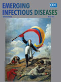
Depictions of Heroism in Battle and Anguish from Tuberculosis [PDF - 332 KB - 2 pages]
| EID | Chorba T, Breedlove B. Depictions of Heroism in Battle and Anguish from Tuberculosis. Emerg Infect Dis. 2016;22(3):573-574. https://doi.org/10.3201/eid2203.ac2203 |
|---|---|
| AMA | Chorba T, Breedlove B. Depictions of Heroism in Battle and Anguish from Tuberculosis. Emerging Infectious Diseases. 2016;22(3):573-574. doi:10.3201/eid2203.ac2203. |
| APA | Chorba, T., & Breedlove, B. (2016). Depictions of Heroism in Battle and Anguish from Tuberculosis. Emerging Infectious Diseases, 22(3), 573-574. https://doi.org/10.3201/eid2203.ac2203. |


