Synopses
Increase in Ocular Syphilis Cases at Ophthalmologic Reference Center, France, 2012–2015 [PDF - 1.50 MB - 8 pages]
We describe the frequency, demographic and clinical features, and visual outcomes of ocular syphilis infections observed during 2012–2015 at a tertiary reference center in Paris, France. Twenty-one cases (29 eyes) were identified. The occurrence of ocular syphilis increased from 1 case in 2012 to 5 cases in 2013, 6 cases in 2014, and 9 cases in 2015 (2.22–25.21/1,000 individual patients/year for the period). Among case-patients, an annual 20%–33% were co-infected with HIV. Seventy-six percent of ocular syphilis infections occurred in men who have sex with men. Seventy-five percent of case-patients had a good final visual outcome (best-corrected visual acuity >0.3 logMAR score). Visual outcome was worse for HIV-positive patients than for HIV-negative patients (p = 0.0139). At follow-up, the best visual outcomes were observed in patients whose mean time from first ocular symptom to consultation was 15 days (SD +19 days).
| EID | Pratas A, Goldschmidt P, Lebeaux D, Aguilar C, Ermak N, Benesty J, et al. Increase in Ocular Syphilis Cases at Ophthalmologic Reference Center, France, 2012–2015. Emerg Infect Dis. 2018;24(2):193-200. https://doi.org/10.3201/eid2402.171167 |
|---|---|
| AMA | Pratas A, Goldschmidt P, Lebeaux D, et al. Increase in Ocular Syphilis Cases at Ophthalmologic Reference Center, France, 2012–2015. Emerging Infectious Diseases. 2018;24(2):193-200. doi:10.3201/eid2402.171167. |
| APA | Pratas, A., Goldschmidt, P., Lebeaux, D., Aguilar, C., Ermak, N., Benesty, J....Errera, M. (2018). Increase in Ocular Syphilis Cases at Ophthalmologic Reference Center, France, 2012–2015. Emerging Infectious Diseases, 24(2), 193-200. https://doi.org/10.3201/eid2402.171167. |
Adenovirus Type 4 Respiratory Infections among Civilian Adults, Northeastern United States, 2011–2015 [PDF - 1.79 MB - 9 pages]
Human adenovirus type 4 (HAdV-4) is most commonly isolated in military settings. We conducted detailed molecular characterization on 36 HAdV-4 isolates recovered from civilian adults with acute respiratory disease (ARD) in the northeastern United States during 2011–2015. Specimens came from college students, residents of long-term care facilities or nursing homes, a cancer patient, and young adults without co-morbidities. HAdV-4 genome types 4a1 and 4a2, the variants most frequently detected among US military recruits in basic training before the restoration of vaccination protocols, were isolated in most cases. Two novel a-like variants were recovered from students enrolled at a college in Tompkins County, New York, USA, and a prototype-like variant distinguishable from the vaccine strain was isolated from an 18-year-old woman visiting a physician’s office in Ulster County, New York, USA, with symptoms of influenza-like illness. Our data suggest that HAdV-4 might be an underestimated causative agent of ARD among civilian adults.
| EID | Kajon AE, Lamson DM, Bair CR, Lu X, Landry ML, Menegus M, et al. Adenovirus Type 4 Respiratory Infections among Civilian Adults, Northeastern United States, 2011–2015. Emerg Infect Dis. 2018;24(2):201-209. https://doi.org/10.3201/eid2402.171407 |
|---|---|
| AMA | Kajon AE, Lamson DM, Bair CR, et al. Adenovirus Type 4 Respiratory Infections among Civilian Adults, Northeastern United States, 2011–2015. Emerging Infectious Diseases. 2018;24(2):201-209. doi:10.3201/eid2402.171407. |
| APA | Kajon, A. E., Lamson, D. M., Bair, C. R., Lu, X., Landry, M. L., Menegus, M....St. George, K. (2018). Adenovirus Type 4 Respiratory Infections among Civilian Adults, Northeastern United States, 2011–2015. Emerging Infectious Diseases, 24(2), 201-209. https://doi.org/10.3201/eid2402.171407. |
Ecologic Features of Plague Outbreak Areas, Democratic Republic of the Congo, 2004–2014 [PDF - 2.91 MB - 11 pages]
During 2004–2014, the Democratic Republic of the Congo (DRC) declared 54% of plague cases worldwide. Using national data, we characterized the epidemiology of human plague in DRC for this period. All 4,630 suspected human plague cases and 349 deaths recorded in DRC came from Orientale Province. Pneumonic plague cases (8.8% of total) occurred during 2 major outbreaks in mining camps in the equatorial forest, and some limited outbreaks occurred in the Ituri highlands. Epidemics originated in 5 health zones clustered in Ituri, where sporadic bubonic cases were recorded throughout every year. Classification and regression tree characterized this cluster by the dominance of ecosystem 40 (mountain tropical climate). In conclusion, a small, stable, endemic focus of plague in the highlands of the Ituri tropical region persisted, acting as a source of outbreaks in DRC.
| EID | Abedi A, Shako J, Gaudart J, Sudre B, Ilunga B, Shamamba S, et al. Ecologic Features of Plague Outbreak Areas, Democratic Republic of the Congo, 2004–2014. Emerg Infect Dis. 2018;24(2):210-220. https://doi.org/10.3201/eid2402.160122 |
|---|---|
| AMA | Abedi A, Shako J, Gaudart J, et al. Ecologic Features of Plague Outbreak Areas, Democratic Republic of the Congo, 2004–2014. Emerging Infectious Diseases. 2018;24(2):210-220. doi:10.3201/eid2402.160122. |
| APA | Abedi, A., Shako, J., Gaudart, J., Sudre, B., Ilunga, B., Shamamba, S....Piarroux, M. (2018). Ecologic Features of Plague Outbreak Areas, Democratic Republic of the Congo, 2004–2014. Emerging Infectious Diseases, 24(2), 210-220. https://doi.org/10.3201/eid2402.160122. |
Liver abscesses containing hypervirulent Klebsiella pneumoniae have emerged during the past 2 decades, originally in Southeast Asia and then worldwide. We hypothesized that hypervirulent K. pneumoniae might also be emerging in France. In a retrospective, monocentric, cohort study, we analyzed characteristics and outcomes for 199 consecutive patients in Paris, France, with liver abscesses during 2010−2015. We focused on 31 patients with abscesses containing K. pneumoniae. This bacterium was present in most (14/27, 52%) cryptogenic liver abscesses. Cryptogenic K. pneumoniae abscesses were more frequently community-acquired (p<0.00001) and monomicrobial (p = 0.008), less likely to involve cancer patients (p<0.01), and relapsed less often (p<0.01) than did noncryptogenic K. pneumoniae liver abscesses. K. pneumoniae isolates from cryptogenic abscesses belonged to either the K1 or K2 serotypes and had more virulence factors than noncryptogenic K. pneumoniae isolates. Hypervirulent K. pneumoniae are emerging as the main pathogen isolated from cryptogenic liver abscesses in the study area.
| EID | Rossi B, Gasperini M, Leflon-Guibout V, Gioanni A, de Lastours V, Rossi G, et al. Hypervirulent Klebsiella pneumoniae in Cryptogenic Liver Abscesses, Paris, France. Emerg Infect Dis. 2018;24(2):221-229. https://doi.org/10.3201/eid2402.170957 |
|---|---|
| AMA | Rossi B, Gasperini M, Leflon-Guibout V, et al. Hypervirulent Klebsiella pneumoniae in Cryptogenic Liver Abscesses, Paris, France. Emerging Infectious Diseases. 2018;24(2):221-229. doi:10.3201/eid2402.170957. |
| APA | Rossi, B., Gasperini, M., Leflon-Guibout, V., Gioanni, A., de Lastours, V., Rossi, G....Lefort, A. (2018). Hypervirulent Klebsiella pneumoniae in Cryptogenic Liver Abscesses, Paris, France. Emerging Infectious Diseases, 24(2), 221-229. https://doi.org/10.3201/eid2402.170957. |
Echinococcus spp. Tapeworms in North America [PDF - 453 KB - 6 pages]
Alveolar and cystic echinococcosis are emerging and reemerging in Europe, Africa, and Asia. The expansion of Echinococcus spp. tapeworms in wildlife host reservoirs appears to be driving this emergence in some areas. Recent studies suggest a similar phenomenon may be occurring in North America. We describe the context of Echinococcus spp. research in North America, with a specific focus on the contiguous United States. Although studies were conducted in the United States throughout the 1900s on various sylvatic and domestic Echinococcus spp. tapeworm cycles, data are lacking for the past ≈30 years. We review previous research, provide analysis of more recent focal studies, and suggest that Echinococcus spp. tapeworms, in particular E. canadensis, may be underrecognized. As a result, we suggest that additional research and surveillance be conducted for these tapeworms in wildlife host reservoirs across the United States.
| EID | Cerda J, Buttke D, Ballweber L. Echinococcus spp. Tapeworms in North America. Emerg Infect Dis. 2018;24(2):230-235. https://doi.org/10.3201/eid2402.161126 |
|---|---|
| AMA | Cerda J, Buttke D, Ballweber L. Echinococcus spp. Tapeworms in North America. Emerging Infectious Diseases. 2018;24(2):230-235. doi:10.3201/eid2402.161126. |
| APA | Cerda, J., Buttke, D., & Ballweber, L. (2018). Echinococcus spp. Tapeworms in North America. Emerging Infectious Diseases, 24(2), 230-235. https://doi.org/10.3201/eid2402.161126. |
Research
Borrelia miyamotoi Infections in Humans and Ticks, Northeastern China [PDF - 1.78 MB - 6 pages]
We conducted an investigation of Borrelia miyamotoi infections in humans and ticks in northeastern China. Of 984 patients reporting recent tick bites, 14 (1.4%) were found to be infected with B. miyamotoi by PCR and genomic sequencing. The 14 patients had nonspecific febrile manifestations, including fever, headache, anorexia, asthenia, and arthralgia. Rash, eschar, and regional lymphadenopathy were each observed in 1 patient. Four (28.6%) patients were hospitalized because of severe disease. B. miyamotoi was detected in 3.0% (19/627) of Ixodes persulcatus, 1 (2.8%) of 36 Haemaphysalis concinna, and none of 29 Dermacentor silvarum ticks. Phylogenetic analyses based on sequences of a nearly entire 16s rRNA gene, a partial flagellin gene, and the glycerophosphodiester phosphodiesterase gene revealed that B. miyamotoi identified in patients and ticks were clustered in the group of the Siberian type. These findings indicate that B. miyamotoi is endemic in northeastern China and its public health significance deserves further investigation.
| EID | Jiang B, Jia N, Jiang J, Zheng Y, Chu Y, Jiang R, et al. Borrelia miyamotoi Infections in Humans and Ticks, Northeastern China. Emerg Infect Dis. 2018;24(2):236-241. https://doi.org/10.3201/eid2402.160378 |
|---|---|
| AMA | Jiang B, Jia N, Jiang J, et al. Borrelia miyamotoi Infections in Humans and Ticks, Northeastern China. Emerging Infectious Diseases. 2018;24(2):236-241. doi:10.3201/eid2402.160378. |
| APA | Jiang, B., Jia, N., Jiang, J., Zheng, Y., Chu, Y., Jiang, R....Fang, L. (2018). Borrelia miyamotoi Infections in Humans and Ticks, Northeastern China. Emerging Infectious Diseases, 24(2), 236-241. https://doi.org/10.3201/eid2402.160378. |
Plasmid-Encoded Transferable mecB-Mediated Methicillin Resistance in Staphylococcus aureus [PDF - 1016 KB - 7 pages]
During cefoxitin-based nasal screening, phenotypically categorized methicillin-resistant Staphylococcus aureus (MRSA) was isolated and tested negative for the presence of the mecA and mecC genes as well as for the SCCmec-orfX junction region. The isolate was found to carry a mecB gene previously described for Macrococcus caseolyticus but not for staphylococcal species. The gene is flanked by β-lactam regulatory genes similar to mecR, mecI, and blaZ and is part of an 84.6-kb multidrug-resistance plasmid that harbors genes encoding additional resistances to aminoglycosides (aacA-aphD, aphA, and aadK) as well as macrolides (ermB) and tetracyclines (tetS). This further plasmidborne β-lactam resistance mechanism harbors the putative risk of acceleration or reacceleration of MRSA spread, resulting in broad ineffectiveness of β-lactams as a main therapeutic application against staphylococcal infections.
| EID | Becker K, van Alen S, Idelevich EA, Schleimer N, Seggewiß J, Mellmann A, et al. Plasmid-Encoded Transferable mecB-Mediated Methicillin Resistance in Staphylococcus aureus. Emerg Infect Dis. 2018;24(2):242-248. https://doi.org/10.3201/eid2402.171074 |
|---|---|
| AMA | Becker K, van Alen S, Idelevich EA, et al. Plasmid-Encoded Transferable mecB-Mediated Methicillin Resistance in Staphylococcus aureus. Emerging Infectious Diseases. 2018;24(2):242-248. doi:10.3201/eid2402.171074. |
| APA | Becker, K., van Alen, S., Idelevich, E. A., Schleimer, N., Seggewiß, J., Mellmann, A....Peters, G. (2018). Plasmid-Encoded Transferable mecB-Mediated Methicillin Resistance in Staphylococcus aureus. Emerging Infectious Diseases, 24(2), 242-248. https://doi.org/10.3201/eid2402.171074. |
Multiplex PCR−Based Next-Generation Sequencing and Global Diversity of Seoul Virus in Humans and Rats [PDF - 1.81 MB - 9 pages]
Seoul virus (SEOV) poses a worldwide public health threat. This virus, which is harbored by Rattus norvegicus and R. rattus rats, is the causative agent of hemorrhagic fever with renal syndrome (HFRS) in humans, which has been reported in Asia, Europe, the Americas, and Africa. Defining SEOV genome sequences plays a critical role in development of preventive and therapeutic strategies against the unique worldwide hantavirus. We applied multiplex PCR–based next-generation sequencing to obtain SEOV genome sequences from clinical and reservoir host specimens. Epidemiologic surveillance of R. norvegicus rats in South Korea during 2000–2016 demonstrated that the serologic prevalence of enzootic SEOV infections was not significant on the basis of sex, weight (age), and season. Viral loads of SEOV in rats showed wide dissemination in tissues and dynamic circulation among populations. Phylogenetic analyses showed the global diversity of SEOV and possible genomic configuration of genetic exchanges.
| EID | Kim W, No J, Lee S, Song D, Lee D, Kim J, et al. Multiplex PCR−Based Next-Generation Sequencing and Global Diversity of Seoul Virus in Humans and Rats. Emerg Infect Dis. 2018;24(2):249-257. https://doi.org/10.3201/eid2402.171216 |
|---|---|
| AMA | Kim W, No J, Lee S, et al. Multiplex PCR−Based Next-Generation Sequencing and Global Diversity of Seoul Virus in Humans and Rats. Emerging Infectious Diseases. 2018;24(2):249-257. doi:10.3201/eid2402.171216. |
| APA | Kim, W., No, J., Lee, S., Song, D., Lee, D., Kim, J....Song, J. (2018). Multiplex PCR−Based Next-Generation Sequencing and Global Diversity of Seoul Virus in Humans and Rats. Emerging Infectious Diseases, 24(2), 249-257. https://doi.org/10.3201/eid2402.171216. |
Staphylococcal toxic shock syndrome (TSS) was originally described in menstruating women and linked to TSS toxin 1 (TSST-1)–producing Staphylococcus aureus. Using UK national surveillance data, we ascertained clinical, molecular and superantigenic characteristics of TSS cases. Average annual TSS incidence was 0.07/100,000 population. Patients with nonmenstrual TSS were younger than those with menstrual TSS but had the same mortality rate. Children <16 years of age accounted for 39% of TSS cases, most caused by burns and skin and soft tissue infections. Nonmenstrual TSS is now more common than menstrual TSS in the UK, although both types are strongly associated with the tst+ clonal complex (CC) 30 methicillin-sensitive S. aureus lineage, which accounted for 49.4% of all TSS and produced more TSST-1 and superantigen bioactivity than did tst+ CC30 methicillin-resistant S. aureus strains. Better understanding of this MSSA lineage and infections in children could focus interventions to prevent TSS in the future.
| EID | Sharma H, Smith D, Turner CE, Game L, Pichon B, Hope R, et al. Clinical and Molecular Epidemiology of Staphylococcal Toxic Shock Syndrome in the United Kingdom. Emerg Infect Dis. 2018;24(2):258-266. https://doi.org/10.3201/eid2402.170606 |
|---|---|
| AMA | Sharma H, Smith D, Turner CE, et al. Clinical and Molecular Epidemiology of Staphylococcal Toxic Shock Syndrome in the United Kingdom. Emerging Infectious Diseases. 2018;24(2):258-266. doi:10.3201/eid2402.170606. |
| APA | Sharma, H., Smith, D., Turner, C. E., Game, L., Pichon, B., Hope, R....Sriskandan, S. (2018). Clinical and Molecular Epidemiology of Staphylococcal Toxic Shock Syndrome in the United Kingdom. Emerging Infectious Diseases, 24(2), 258-266. https://doi.org/10.3201/eid2402.170606. |
Lethal Respiratory Disease Associated with Human Rhinovirus C in Wild Chimpanzees, Uganda, 2013 [PDF - 1.52 MB - 8 pages]
We describe a lethal respiratory outbreak among wild chimpanzees in Uganda in 2013 for which molecular and epidemiologic analyses implicate human rhinovirus C as the cause. Postmortem samples from an infant chimpanzee yielded near-complete genome sequences throughout the respiratory tract; other pathogens were absent. Epidemiologic modeling estimated the basic reproductive number (R0) for the epidemic as 1.83, consistent with the common cold in humans. Genotyping of 41 chimpanzees and examination of 24 published chimpanzee genomes from subspecies across Africa showed universal homozygosity for the cadherin-related family member 3 CDHR3-Y529 allele, which increases risk for rhinovirus C infection and asthma in human children. These results indicate that chimpanzees exhibit a species-wide genetic susceptibility to rhinovirus C and that this virus, heretofore considered a uniquely human pathogen, can cross primate species barriers and threatens wild apes. We advocate engineering interventions and prevention strategies for rhinovirus infections for both humans and wild apes.
| EID | Scully EJ, Basnet S, Wrangham RW, Muller MN, Otali E, Hyeroba D, et al. Lethal Respiratory Disease Associated with Human Rhinovirus C in Wild Chimpanzees, Uganda, 2013. Emerg Infect Dis. 2018;24(2):267-274. https://doi.org/10.3201/eid2402.170778 |
|---|---|
| AMA | Scully EJ, Basnet S, Wrangham RW, et al. Lethal Respiratory Disease Associated with Human Rhinovirus C in Wild Chimpanzees, Uganda, 2013. Emerging Infectious Diseases. 2018;24(2):267-274. doi:10.3201/eid2402.170778. |
| APA | Scully, E. J., Basnet, S., Wrangham, R. W., Muller, M. N., Otali, E., Hyeroba, D....Goldberg, T. L. (2018). Lethal Respiratory Disease Associated with Human Rhinovirus C in Wild Chimpanzees, Uganda, 2013. Emerging Infectious Diseases, 24(2), 267-274. https://doi.org/10.3201/eid2402.170778. |
Spread of Meropenem-Resistant Streptococcus pneumoniae Serotype 15A-ST63 Clone in Japan, 2012–2014 [PDF - 1.86 MB - 9 pages]
After the introduction of pneumococcal conjugate vaccines, the incidence of pneumococcal infections due to meropenem-resistant serotype 15A-ST63 strains increased in Japan. By using whole-genome sequencing and comparing sequences with those of clones from the United Kingdom, the United States, and Canada, we clarified the traits of the serotype 15A-ST63 clone. Our analysis revealed that the meropenem-resistant serotype 15A-ST63 strains from Japan originated from meropenem-susceptible strains from Japan. Recombination site prediction analysis showed that the meropenem-resistant strain-specific recombination regions included the pbp1a and pbp2b regions. A detailed analysis of the composition of these genes indicated that resistance seems to be caused by pbp1a recombination. The pbp1a gene in meropenem-resistant isolates was identical to that in multidrug (including meropenem)–resistant serotype 19A-ST320 pneumococci, which have spread in the United States. The global spread of pneumococci of this lineage is noteworthy because serotype 15A is not included in the currently used 13-valent pneumococcal conjugate vaccine.
| EID | Nakano S, Fujisawa T, Ito Y, Chang B, Matsumura Y, Yamamoto M, et al. Spread of Meropenem-Resistant Streptococcus pneumoniae Serotype 15A-ST63 Clone in Japan, 2012–2014. Emerg Infect Dis. 2018;24(2):275-283. https://doi.org/10.3201/eid2402.171268 |
|---|---|
| AMA | Nakano S, Fujisawa T, Ito Y, et al. Spread of Meropenem-Resistant Streptococcus pneumoniae Serotype 15A-ST63 Clone in Japan, 2012–2014. Emerging Infectious Diseases. 2018;24(2):275-283. doi:10.3201/eid2402.171268. |
| APA | Nakano, S., Fujisawa, T., Ito, Y., Chang, B., Matsumura, Y., Yamamoto, M....Ichiyama, S. (2018). Spread of Meropenem-Resistant Streptococcus pneumoniae Serotype 15A-ST63 Clone in Japan, 2012–2014. Emerging Infectious Diseases, 24(2), 275-283. https://doi.org/10.3201/eid2402.171268. |
Role of Environmental Factors in Shaping Spatial Distribution of Salmonella enterica Serovar Typhi, Fiji [PDF - 2.12 MB - 10 pages]
Fiji recently experienced a sharp increase in reported typhoid fever cases. To investigate geographic distribution and environmental risk factors associated with Salmonella enterica serovar Typhi infection, we conducted a cross-sectional cluster survey with associated serologic testing for Vi capsular antigen–specific antibodies (a marker for exposure to Salmonella Typhi in Fiji in 2013. Hotspots with high seroprevalence of Vi-specific antibodies were identified in northeastern mainland Fiji. Risk for Vi seropositivity increased with increased annual rainfall (odds ratio [OR] 1.26/quintile increase, 95% CI 1.12–1.42), and decreased with increased distance from major rivers and creeks (OR 0.89/km increase, 95% CI 0.80–0.99) and distance to modeled flood-risk areas (OR 0.80/quintile increase, 95% CI 0.69–0.92) after being adjusted for age, typhoid fever vaccination, and home toilet type. Risk for exposure to Salmonella Typhi and its spatial distribution in Fiji are driven by environmental factors. Our findings can directly affect typhoid fever control efforts in Fiji.
| EID | de Alwis R, Watson C, Nikolay B, Lowry JH, Thieu N, Van T, et al. Role of Environmental Factors in Shaping Spatial Distribution of Salmonella enterica Serovar Typhi, Fiji. Emerg Infect Dis. 2018;24(2):284-293. https://doi.org/10.3201/eid2402.170704 |
|---|---|
| AMA | de Alwis R, Watson C, Nikolay B, et al. Role of Environmental Factors in Shaping Spatial Distribution of Salmonella enterica Serovar Typhi, Fiji. Emerging Infectious Diseases. 2018;24(2):284-293. doi:10.3201/eid2402.170704. |
| APA | de Alwis, R., Watson, C., Nikolay, B., Lowry, J. H., Thieu, N., Van, T....Cano, J. (2018). Role of Environmental Factors in Shaping Spatial Distribution of Salmonella enterica Serovar Typhi, Fiji. Emerging Infectious Diseases, 24(2), 284-293. https://doi.org/10.3201/eid2402.170704. |
Yersinia pestis Survival and Replication in Potential Ameba Reservoir [PDF - 2.08 MB - 9 pages]
Plague ecology is characterized by sporadic epizootics, then periods of dormancy. Building evidence suggests environmentally ubiquitous amebae act as feral macrophages and hosts to many intracellular pathogens. We conducted environmental genetic surveys and laboratory co-culture infection experiments to assess whether plague bacteria were resistant to digestion by 5 environmental ameba species. First, we demonstrated that Yersinia pestis is resistant or transiently resistant to various ameba species. Second, we showed that Y. pestis survives and replicates intracellularly within Dictyostelium discoideum amebae for ˃48 hours postinfection, whereas control bacteria were destroyed in <1 hour. Finally, we found that Y. pestis resides within ameba structures synonymous with those found in infected human macrophages, for which Y. pestis is a competent pathogen. Evidence supporting amebae as potential plague reservoirs stresses the importance of recognizing pathogen-harboring amebae as threats to public health, agriculture, conservation, and biodefense.
| EID | Markman DW, Antolin MF, Bowen RA, Wheat WH, Woods M, Gonzalez-Juarrero M, et al. Yersinia pestis Survival and Replication in Potential Ameba Reservoir. Emerg Infect Dis. 2018;24(2):294-302. https://doi.org/10.3201/eid2402.171065 |
|---|---|
| AMA | Markman DW, Antolin MF, Bowen RA, et al. Yersinia pestis Survival and Replication in Potential Ameba Reservoir. Emerging Infectious Diseases. 2018;24(2):294-302. doi:10.3201/eid2402.171065. |
| APA | Markman, D. W., Antolin, M. F., Bowen, R. A., Wheat, W. H., Woods, M., Gonzalez-Juarrero, M....Jackson, M. (2018). Yersinia pestis Survival and Replication in Potential Ameba Reservoir. Emerging Infectious Diseases, 24(2), 294-302. https://doi.org/10.3201/eid2402.171065. |
New Parvovirus Associated with Serum Hepatitis in Horses after Inoculation of Common Biological Product [PDF - 2.00 MB - 8 pages]
Equine serum hepatitis (i.e., Theiler’s disease) is a serious and often life-threatening disease of unknown etiology that affects horses. A horse in Nebraska, USA, with serum hepatitis died 65 days after treatment with equine-origin tetanus antitoxin. We identified an unknown parvovirus in serum and liver of the dead horse and in the administered antitoxin. The equine parvovirus-hepatitis (EqPV-H) shares <50% protein identity with its phylogenetic relatives of the genus Copiparvovirus. Next, we experimentally infected 2 horses using a tetanus antitoxin contaminated with EqPV-H. Viremia developed, the horses seroconverted, and acute hepatitis developed that was confirmed by clinical, biochemical, and histopathologic testing. We also determined that EqPV-H is an endemic infection because, in a cohort of 100 clinically normal adult horses, 13 were viremic and 15 were seropositive. We identified a new virus associated with equine serum hepatitis and confirmed its pathogenicity and transmissibility through contaminated biological products.
| EID | Divers TJ, Tennant BC, Kumar A, McDonough S, Cullen J, Bhuva N, et al. New Parvovirus Associated with Serum Hepatitis in Horses after Inoculation of Common Biological Product. Emerg Infect Dis. 2018;24(2):303-310. https://doi.org/10.3201/eid2402.171031 |
|---|---|
| AMA | Divers TJ, Tennant BC, Kumar A, et al. New Parvovirus Associated with Serum Hepatitis in Horses after Inoculation of Common Biological Product. Emerging Infectious Diseases. 2018;24(2):303-310. doi:10.3201/eid2402.171031. |
| APA | Divers, T. J., Tennant, B. C., Kumar, A., McDonough, S., Cullen, J., Bhuva, N....Kapoor, A. (2018). New Parvovirus Associated with Serum Hepatitis in Horses after Inoculation of Common Biological Product. Emerging Infectious Diseases, 24(2), 303-310. https://doi.org/10.3201/eid2402.171031. |
Development of a Pediatric Ebola Predictive Score, Sierra Leone [PDF - 1.01 MB - 9 pages]
We compared children who were positive for Ebola virus disease (EVD) with those who were negative to derive a pediatric EVD predictor (PEP) score. We collected data on all children <13 years of age admitted to 11 Ebola holding units in Sierra Leone during August 2014–March 2015 and performed multivariable logistic regression. Among 1,054 children, 309 (29%) were EVD positive and 697 (66%) EVD negative, with 48 (5%) missing. Contact history, conjunctivitis, and age were the strongest positive predictors for EVD. The PEP score had an area under receiver operating characteristics curve of 0.80. A PEP score of 7/10 was 92% specific and 44% sensitive; 3/10 was 30% specific, 94% sensitive. The PEP score could correctly classify 79%–90% of children and could be used to facilitate triage into risk categories, depending on the sensitivity or specificity required.
| EID | Fitzgerald F, Wing K, Naveed A, Gbessay M, Ross J, Checchi F, et al. Development of a Pediatric Ebola Predictive Score, Sierra Leone. Emerg Infect Dis. 2018;24(2):311-319. https://doi.org/10.3201/eid2402.171018 |
|---|---|
| AMA | Fitzgerald F, Wing K, Naveed A, et al. Development of a Pediatric Ebola Predictive Score, Sierra Leone. Emerging Infectious Diseases. 2018;24(2):311-319. doi:10.3201/eid2402.171018. |
| APA | Fitzgerald, F., Wing, K., Naveed, A., Gbessay, M., Ross, J., Checchi, F....Yeung, S. (2018). Development of a Pediatric Ebola Predictive Score, Sierra Leone. Emerging Infectious Diseases, 24(2), 311-319. https://doi.org/10.3201/eid2402.171018. |
Trends in Infectious Disease Mortality, South Korea, 1983–2015 [PDF - 811 KB - 8 pages]
We used national statistics from 1983–2015 to evaluate trends in mortality caused by infectious diseases in South Korea. Age-standardized mortality from infectious disease decreased from 43.5/100,000 population in 1983 to 16.5/100,000 in 1996, and then increased to 44.6/100,000 in 2015. Tuberculosis was the most common cause of death in 1983 and respiratory tract infections in 2015. We observed a significant decline in infant deaths caused by infectious diseases, but mortality in persons age >65 years increased from 135 deaths/100,000 population in 1996 to 307/100,000 in 2015. The relative inequality indices for respiratory tract infections, sepsis, and tuberculosis tended to increase over time. Although substantial progress has been achieved in terms of infant mortality, death rates from infectious disease has not decreased overall. Elderly populations with lower education levels and subgroups susceptible to respiratory infections and sepsis should be the focus of preventive policies.
| EID | Choe Y, Choe S, Cho S. Trends in Infectious Disease Mortality, South Korea, 1983–2015. Emerg Infect Dis. 2018;24(2):320-327. https://doi.org/10.3201/eid2402.170862 |
|---|---|
| AMA | Choe Y, Choe S, Cho S. Trends in Infectious Disease Mortality, South Korea, 1983–2015. Emerging Infectious Diseases. 2018;24(2):320-327. doi:10.3201/eid2402.170862. |
| APA | Choe, Y., Choe, S., & Cho, S. (2018). Trends in Infectious Disease Mortality, South Korea, 1983–2015. Emerging Infectious Diseases, 24(2), 320-327. https://doi.org/10.3201/eid2402.170862. |
Use of Pristinamycin for Macrolide-Resistant Mycoplasma genitalium Infection [PDF - 623 KB - 8 pages]
High levels of macrolide resistance and increasing fluoroquinolone resistance are found in Mycoplasma genitalium in many countries. We evaluated pristinamycin for macrolide-resistant M. genitalium in a sexual health center in Australia. Microbiologic cure was determined by M. genitalium–specific 16S PCR 14–90 days after treatment began. Of 114 persons treated with pristinamycin, infection was cured in 85 (75%). This percentage did not change when pristinamycin was given at daily doses of 2 g or 4 g or at 3 g combined with 200 mg doxycycline. In infections with higher pretreatment bacterial load, treatment was twice as likely to fail for each 1 log10 increase in bacterial load. Gastrointestinal side effects occurred in 7% of patients. Pristinamycin at maximum oral dose, or combined with doxycycline, cured 75% of macrolide-resistant M. genitalium infections. Pristinamycin is well-tolerated and remains an option where fluoroquinolones have failed or cannot be used.
| EID | Read T, Jensen JS, Fairley CK, Grant M, Danielewski JA, Su J, et al. Use of Pristinamycin for Macrolide-Resistant Mycoplasma genitalium Infection. Emerg Infect Dis. 2018;24(2):328-335. https://doi.org/10.3201/eid2402.170902 |
|---|---|
| AMA | Read T, Jensen JS, Fairley CK, et al. Use of Pristinamycin for Macrolide-Resistant Mycoplasma genitalium Infection. Emerging Infectious Diseases. 2018;24(2):328-335. doi:10.3201/eid2402.170902. |
| APA | Read, T., Jensen, J. S., Fairley, C. K., Grant, M., Danielewski, J. A., Su, J....Bradshaw, C. S. (2018). Use of Pristinamycin for Macrolide-Resistant Mycoplasma genitalium Infection. Emerging Infectious Diseases, 24(2), 328-335. https://doi.org/10.3201/eid2402.170902. |
Risk Communication and Ebola-Specific Knowledge and Behavior during 2014–2015 Outbreak, Sierra Leone [PDF - 510 KB - 9 pages]
We assessed the effect of information sources on Ebola-specific knowledge and behavior during the 2014–2015 Ebola virus disease outbreak in Sierra Leone. We pooled data from 4 population-based knowledge, attitude, and practice surveys (August, October, and December 2014 and July 2015), with a total of 10,604 respondents. We created composite variables for exposures (information sources: electronic, print, new media, government, community) and outcomes (knowledge and misconceptions, protective and risk behavior) and tested associations by using logistic regression within multilevel modeling. Exposure to information sources was associated with higher knowledge and protective behaviors. However, apart from print media, exposure to information sources was also linked to misconceptions and risk behavior, but with weaker associations observed. Knowledge and protective behavior were associated with the outbreak level, most strongly after the peak, whereas risk behavior was seen at all levels of the outbreak. In future outbreaks, close attention should be paid to dissemination of information.
| EID | Winters M, Jalloh MF, Sengeh P, Jalloh MB, Conteh L, Bunnell R, et al. Risk Communication and Ebola-Specific Knowledge and Behavior during 2014–2015 Outbreak, Sierra Leone. Emerg Infect Dis. 2018;24(2):336-344. https://doi.org/10.3201/eid2402.171028 |
|---|---|
| AMA | Winters M, Jalloh MF, Sengeh P, et al. Risk Communication and Ebola-Specific Knowledge and Behavior during 2014–2015 Outbreak, Sierra Leone. Emerging Infectious Diseases. 2018;24(2):336-344. doi:10.3201/eid2402.171028. |
| APA | Winters, M., Jalloh, M. F., Sengeh, P., Jalloh, M. B., Conteh, L., Bunnell, R....Nordenstedt, H. (2018). Risk Communication and Ebola-Specific Knowledge and Behavior during 2014–2015 Outbreak, Sierra Leone. Emerging Infectious Diseases, 24(2), 336-344. https://doi.org/10.3201/eid2402.171028. |
Macacine Herpesvirus 1 Antibody Prevalence and DNA Shedding among Invasive Rhesus Macaques, Silver Springs State Park, Florida, USA [PDF - 604 KB - 7 pages]
We compiled records on macacine herpesvirus 1 (McHV-1) seroprevalence and, during 2015–2016, collected saliva and fecal samples from the free-ranging rhesus macaques of Silver Springs State Park, a popular public park in central Florida, USA, to determine viral DNA shedding and perform sequencing. Phylogenetic analysis of the US5 and US5-US6 intragenic sequence from free-ranging and laboratory McHV-1 variants did not reveal genomic differences. In animals captured during 2000–2012, average annual seroprevalence was 25% ± 9 (mean ± SD). We found 4%–14% (95% CI 2%–29%) of macaques passively sampled during the fall 2015 mating season shed McHV-1 DNA orally. We did not observe viral shedding during the spring or summer or from fecal samples. We conclude that these macaques can shed McHV-1, putting humans at risk for exposure to this potentially fatal pathogen. Management plans should be put in place to limit transmission of McHV-1 from these macaques.
| EID | Wisely SM, Sayler KA, Anderson C, Boyce CL, Klegarth AR, Johnson SA. Macacine Herpesvirus 1 Antibody Prevalence and DNA Shedding among Invasive Rhesus Macaques, Silver Springs State Park, Florida, USA. Emerg Infect Dis. 2018;24(2):345-351. https://doi.org/10.3201/eid2402.171439 |
|---|---|
| AMA | Wisely SM, Sayler KA, Anderson C, et al. Macacine Herpesvirus 1 Antibody Prevalence and DNA Shedding among Invasive Rhesus Macaques, Silver Springs State Park, Florida, USA. Emerging Infectious Diseases. 2018;24(2):345-351. doi:10.3201/eid2402.171439. |
| APA | Wisely, S. M., Sayler, K. A., Anderson, C., Boyce, C. L., Klegarth, A. R., & Johnson, S. A. (2018). Macacine Herpesvirus 1 Antibody Prevalence and DNA Shedding among Invasive Rhesus Macaques, Silver Springs State Park, Florida, USA. Emerging Infectious Diseases, 24(2), 345-351. https://doi.org/10.3201/eid2402.171439. |
Dispatches
Co-circulation of Influenza A H5, H7, and H9 Viruses and Co-infected Poultry in Live Bird Markets, Cambodia [PDF - 1.38 MB - 4 pages]
Longitudinal surveillance of 2 live bird markets in Cambodia revealed year-round, high co-circulation of H5, H7, and H9 influenza viruses. We detected influenza A viruses in 51.3% of ducks and 39.6% of chickens, and co-infections, mainly by H5 and H9 viruses, in 0.8% of ducks and 4.5% of chickens.
| EID | Horwood PF, Horm S, Suttie A, Thet S, Y P, Rith S, et al. Co-circulation of Influenza A H5, H7, and H9 Viruses and Co-infected Poultry in Live Bird Markets, Cambodia. Emerg Infect Dis. 2018;24(2):352-355. https://doi.org/10.3201/eid2402.171360 |
|---|---|
| AMA | Horwood PF, Horm S, Suttie A, et al. Co-circulation of Influenza A H5, H7, and H9 Viruses and Co-infected Poultry in Live Bird Markets, Cambodia. Emerging Infectious Diseases. 2018;24(2):352-355. doi:10.3201/eid2402.171360. |
| APA | Horwood, P. F., Horm, S., Suttie, A., Thet, S., Y, P., Rith, S....Dussart, P. (2018). Co-circulation of Influenza A H5, H7, and H9 Viruses and Co-infected Poultry in Live Bird Markets, Cambodia. Emerging Infectious Diseases, 24(2), 352-355. https://doi.org/10.3201/eid2402.171360. |
Effects of Culling on Leptospira interrogans Carriage by Rats [PDF - 2.62 MB - 5 pages]
We found that lethal, urban rat control is associated with a significant increase in the odds that surviving rats carry Leptospira interrogans. Our results suggest that human interventions have the potential to affect and even increase the prevalence of zoonotic pathogens within rat populations.
| EID | Lee MJ, Byers KA, Donovan CM, Bidulka JJ, Stephen C, Patrick DM, et al. Effects of Culling on Leptospira interrogans Carriage by Rats. Emerg Infect Dis. 2018;24(2):356-360. https://doi.org/10.3201/eid2402.171371 |
|---|---|
| AMA | Lee MJ, Byers KA, Donovan CM, et al. Effects of Culling on Leptospira interrogans Carriage by Rats. Emerging Infectious Diseases. 2018;24(2):356-360. doi:10.3201/eid2402.171371. |
| APA | Lee, M. J., Byers, K. A., Donovan, C. M., Bidulka, J. J., Stephen, C., Patrick, D. M....Himsworth, C. G. (2018). Effects of Culling on Leptospira interrogans Carriage by Rats. Emerging Infectious Diseases, 24(2), 356-360. https://doi.org/10.3201/eid2402.171371. |
Scrub Typhus Outbreak in Chonburi Province, Central Thailand, 2013 [PDF - 2.17 MB - 5 pages]
Investigation of a scrub typhus outbreak in Thailand during September 2013 found that 9.1% of Thai soldiers and 11.1% of residents living in areas surrounding training sites had antibodies against the causative agent, Orientia tsutsugamushi. Sequence analysis of O. tsutsugamushi from rodents and chiggers identified 7 genogroups and 3 genotypes.
| EID | Rodkvamtook W, Kuttasingkee N, Linsuwanon P, Sudsawat Y, Richards AL, Somsri M, et al. Scrub Typhus Outbreak in Chonburi Province, Central Thailand, 2013. Emerg Infect Dis. 2018;24(2):361-365. https://doi.org/10.3201/eid2402.171172 |
|---|---|
| AMA | Rodkvamtook W, Kuttasingkee N, Linsuwanon P, et al. Scrub Typhus Outbreak in Chonburi Province, Central Thailand, 2013. Emerging Infectious Diseases. 2018;24(2):361-365. doi:10.3201/eid2402.171172. |
| APA | Rodkvamtook, W., Kuttasingkee, N., Linsuwanon, P., Sudsawat, Y., Richards, A. L., Somsri, M....Gaywee, J. (2018). Scrub Typhus Outbreak in Chonburi Province, Central Thailand, 2013. Emerging Infectious Diseases, 24(2), 361-365. https://doi.org/10.3201/eid2402.171172. |
Epidemic Varicella Zoster Virus among University Students, India [PDF - 656 KB - 4 pages]
We investigated a yearlong varicella zoster virus outbreak in a highly susceptible young adult population at a large university in India. Outbreaks of varicella infection among adults are not well described in the literature. Infection control measures and vaccination policy for this age group and setting are needed.
| EID | Meyers J, Logaraj M, Ramraj B, Narasimhan P, MacIntyre C. Epidemic Varicella Zoster Virus among University Students, India. Emerg Infect Dis. 2018;24(2):366-369. https://doi.org/10.3201/eid2402.170659 |
|---|---|
| AMA | Meyers J, Logaraj M, Ramraj B, et al. Epidemic Varicella Zoster Virus among University Students, India. Emerging Infectious Diseases. 2018;24(2):366-369. doi:10.3201/eid2402.170659. |
| APA | Meyers, J., Logaraj, M., Ramraj, B., Narasimhan, P., & MacIntyre, C. (2018). Epidemic Varicella Zoster Virus among University Students, India. Emerging Infectious Diseases, 24(2), 366-369. https://doi.org/10.3201/eid2402.170659. |
Fly Reservoir Associated with Wohlfahrtiimonas Bacteremia in a Human [PDF - 1.30 MB - 4 pages]
Wohlfahrtiimonas species bacteria were isolated from the bloodstream of a patient with septicemia and wound myiasis. Environmental investigations identified a Wohlfahrtiimonas sp. among insects in the Americas and in a previously undescribed vector, the green bottle fly (Lucilia sericata). The isolates possibly represent a new species within the genus Wohlfahrtiimonas.
| EID | Bonwitt JH, Tran M, Dykstra EA, Eckmann K, Bell ME, Leadon M, et al. Fly Reservoir Associated with Wohlfahrtiimonas Bacteremia in a Human. Emerg Infect Dis. 2018;24(2):370-373. https://doi.org/10.3201/eid2402.170913 |
|---|---|
| AMA | Bonwitt JH, Tran M, Dykstra EA, et al. Fly Reservoir Associated with Wohlfahrtiimonas Bacteremia in a Human. Emerging Infectious Diseases. 2018;24(2):370-373. doi:10.3201/eid2402.170913. |
| APA | Bonwitt, J. H., Tran, M., Dykstra, E. A., Eckmann, K., Bell, M. E., Leadon, M....Glover, W. A. (2018). Fly Reservoir Associated with Wohlfahrtiimonas Bacteremia in a Human. Emerging Infectious Diseases, 24(2), 370-373. https://doi.org/10.3201/eid2402.170913. |
Containment of Highly Pathogenic Avian Influenza A(H5N1) Virus, Lebanon, 2016 [PDF - 1.07 MB - 3 pages]
A preparedness plan for avian influenza A(H5N1) virus infection was activated in Lebanon in 2016 after reported cases in poultry. Exposed persons were given prophylaxis and monitored daily. A total of 185 exposed persons were identified: 180 received prophylaxis, 181 were monitored, and 41 suspected cases were reported. All collected specimens were negative for virus by PCR.
| EID | Farah ZE, Khatib O, Hamadeh S, Ahmad K, El Bazzal B, Zalloua P, et al. Containment of Highly Pathogenic Avian Influenza A(H5N1) Virus, Lebanon, 2016. Emerg Infect Dis. 2018;24(2):374-376. https://doi.org/10.3201/eid2402.171276 |
|---|---|
| AMA | Farah ZE, Khatib O, Hamadeh S, et al. Containment of Highly Pathogenic Avian Influenza A(H5N1) Virus, Lebanon, 2016. Emerging Infectious Diseases. 2018;24(2):374-376. doi:10.3201/eid2402.171276. |
| APA | Farah, Z. E., Khatib, O., Hamadeh, S., Ahmad, K., El Bazzal, B., Zalloua, P....Ghosn, N. (2018). Containment of Highly Pathogenic Avian Influenza A(H5N1) Virus, Lebanon, 2016. Emerging Infectious Diseases, 24(2), 374-376. https://doi.org/10.3201/eid2402.171276. |
Emergomyces africanus in Soil, South Africa [PDF - 893 KB - 4 pages]
We detected Emergomyces africanus, a thermally dimorphic fungus that causes an HIV-associated systemic mycosis, by PCR in 18 (30%) of 60 soil samples from a wide range of habitats in South Africa. Direct and indirect culture techniques were unsuccessful. Experimental intraperitoneal inoculation of conidia induced murine disease.
| EID | Schwartz IS, Lerm B, Hoving J, Kenyon C, Horsnell WG, Basson W, et al. Emergomyces africanus in Soil, South Africa. Emerg Infect Dis. 2018;24(2):377-380. https://doi.org/10.3201/eid2402.171351 |
|---|---|
| AMA | Schwartz IS, Lerm B, Hoving J, et al. Emergomyces africanus in Soil, South Africa. Emerging Infectious Diseases. 2018;24(2):377-380. doi:10.3201/eid2402.171351. |
| APA | Schwartz, I. S., Lerm, B., Hoving, J., Kenyon, C., Horsnell, W. G., Basson, W....Botha, A. (2018). Emergomyces africanus in Soil, South Africa. Emerging Infectious Diseases, 24(2), 377-380. https://doi.org/10.3201/eid2402.171351. |
Ceftriaxone-Resistant Neisseria gonorrhoeae, Canada, 2017 [PDF - 726 KB - 3 pages]
We identified a ceftriaxone-resistant Neisseria gonorrhoeae isolate in a patient in Canada. This isolate carried the penA-60 allele, which differs substantially from its closest relative, mosaic penA XXVII (80% nucleotide identity). Epidemiologic and genomic data suggest spread from Asia. Antimicrobial susceptibility surveillance helps prevent spread of highly resistant N. gonorrhoeae strains.
| EID | Lefebvre B, Martin I, Demczuk W, Deshaies L, Michaud S, Labbé A, et al. Ceftriaxone-Resistant Neisseria gonorrhoeae, Canada, 2017. Emerg Infect Dis. 2018;24(2):381-383. https://doi.org/10.3201/eid2402.171756 |
|---|---|
| AMA | Lefebvre B, Martin I, Demczuk W, et al. Ceftriaxone-Resistant Neisseria gonorrhoeae, Canada, 2017. Emerging Infectious Diseases. 2018;24(2):381-383. doi:10.3201/eid2402.171756. |
| APA | Lefebvre, B., Martin, I., Demczuk, W., Deshaies, L., Michaud, S., Labbé, A....Longtin, J. (2018). Ceftriaxone-Resistant Neisseria gonorrhoeae, Canada, 2017. Emerging Infectious Diseases, 24(2), 381-383. https://doi.org/10.3201/eid2402.171756. |
Clusters of Human Infection and Human-to-Human Transmission of Avian Influenza A(H7N9) Virus, 2013–2017 [PDF - 385 KB - 4 pages]
To detect changes in human-to-human transmission of influenza A(H7N9) virus, we analyzed characteristics of 40 clusters of case-patients during 5 epidemics in China in 2013–2017. Similarities in number and size of clusters and proportion of clusters with probable human-to-human transmission across all epidemics suggest no change in human-to-human transmission risk.
| EID | Zhou L, Chen E, Bao C, Xiang N, Wu J, Wu S, et al. Clusters of Human Infection and Human-to-Human Transmission of Avian Influenza A(H7N9) Virus, 2013–2017. Emerg Infect Dis. 2018;24(2):397-400. https://doi.org/10.3201/eid2402.171565 |
|---|---|
| AMA | Zhou L, Chen E, Bao C, et al. Clusters of Human Infection and Human-to-Human Transmission of Avian Influenza A(H7N9) Virus, 2013–2017. Emerging Infectious Diseases. 2018;24(2):397-400. doi:10.3201/eid2402.171565. |
| APA | Zhou, L., Chen, E., Bao, C., Xiang, N., Wu, J., Wu, S....Li, Q. (2018). Clusters of Human Infection and Human-to-Human Transmission of Avian Influenza A(H7N9) Virus, 2013–2017. Emerging Infectious Diseases, 24(2), 397-400. https://doi.org/10.3201/eid2402.171565. |
Research Letters
Cysticercosis in Shandong Province, Eastern China [PDF - 332 KB - 2 pages]
We analyzed demographic and clinical data and estimated the incidence of cysticercosis in Shandong Province, China, during 1975–2014. Our analyses showed that a cysticercosis-endemic area is present in Shandong Province, especially in its western regions. Improved surveillance and control are needed to address the elevated risk for cysticercosis in this region.
| EID | Liu G, Li Y, Cui Y, Huang B, Wang H, Chen Y, et al. Cysticercosis in Shandong Province, Eastern China. Emerg Infect Dis. 2018;24(2):384-385. https://doi.org/10.3201/eid2402.151253 |
|---|---|
| AMA | Liu G, Li Y, Cui Y, et al. Cysticercosis in Shandong Province, Eastern China. Emerging Infectious Diseases. 2018;24(2):384-385. doi:10.3201/eid2402.151253. |
| APA | Liu, G., Li, Y., Cui, Y., Huang, B., Wang, H., Chen, Y....Liu, X. (2018). Cysticercosis in Shandong Province, Eastern China. Emerging Infectious Diseases, 24(2), 384-385. https://doi.org/10.3201/eid2402.151253. |
Rickettsia africae and Novel Rickettsial Strain in Amblyomma spp. Ticks, Nicaragua, 2013 [PDF - 474 KB - 3 pages]
We report molecular detection of Rickettsia africae in Amblyomma ovale ticks from Nicaragua and a novel rickettsial strain in an A. triste tick. Of 146 ticks from dogs, 16.4% were Rickettsia PCR positive. The presence of Rickettsia spp. in human-biting ticks in Nicaragua may pose a public health concern.
| EID | Vogel H, Foley J, Fiorello CV. Rickettsia africae and Novel Rickettsial Strain in Amblyomma spp. Ticks, Nicaragua, 2013. Emerg Infect Dis. 2018;24(2):385-387. https://doi.org/10.3201/eid2402.161901 |
|---|---|
| AMA | Vogel H, Foley J, Fiorello CV. Rickettsia africae and Novel Rickettsial Strain in Amblyomma spp. Ticks, Nicaragua, 2013. Emerging Infectious Diseases. 2018;24(2):385-387. doi:10.3201/eid2402.161901. |
| APA | Vogel, H., Foley, J., & Fiorello, C. V. (2018). Rickettsia africae and Novel Rickettsial Strain in Amblyomma spp. Ticks, Nicaragua, 2013. Emerging Infectious Diseases, 24(2), 385-387. https://doi.org/10.3201/eid2402.161901. |
Amebaborne “Attilina massiliensis” Keratitis, France [PDF - 810 KB - 3 pages]
We report a case of Acanthamoeba castellani keratitis in a person who wore contact lenses. The amebae hosted an ameba-resistant bacterial symbiont, provisionally named “Attilina massiliensis,” a yet undescribed α-Proteobacterium.
| EID | Battaini A, La Scola B, Yin G, Hoffart L, Drancourt M. Amebaborne “Attilina massiliensis” Keratitis, France. Emerg Infect Dis. 2018;24(2):387-389. https://doi.org/10.3201/eid2402.170541 |
|---|---|
| AMA | Battaini A, La Scola B, Yin G, et al. Amebaborne “Attilina massiliensis” Keratitis, France. Emerging Infectious Diseases. 2018;24(2):387-389. doi:10.3201/eid2402.170541. |
| APA | Battaini, A., La Scola, B., Yin, G., Hoffart, L., & Drancourt, M. (2018). Amebaborne “Attilina massiliensis” Keratitis, France. Emerging Infectious Diseases, 24(2), 387-389. https://doi.org/10.3201/eid2402.170541. |
Influenza D Virus in Cattle, Ireland [PDF - 363 KB - 3 pages]
We detected influenza D virus in 18 nasal swab samples from cattle in Ireland that were clinically diagnosed with respiratory disease. Specimens were obtained from archived samples received for routine diagnosis during 2014–2016. Sequencing showed that viruses from Ireland clustered with virus sequences obtained in Europe within the D/swine/OK/1334/2011 clade.
| EID | Flynn O, Gallagher C, Mooney J, Irvine C, Ducatez M, Hause B, et al. Influenza D Virus in Cattle, Ireland. Emerg Infect Dis. 2018;24(2):389-391. https://doi.org/10.3201/eid2402.170759 |
|---|---|
| AMA | Flynn O, Gallagher C, Mooney J, et al. Influenza D Virus in Cattle, Ireland. Emerging Infectious Diseases. 2018;24(2):389-391. doi:10.3201/eid2402.170759. |
| APA | Flynn, O., Gallagher, C., Mooney, J., Irvine, C., Ducatez, M., Hause, B....Ryan, E. (2018). Influenza D Virus in Cattle, Ireland. Emerging Infectious Diseases, 24(2), 389-391. https://doi.org/10.3201/eid2402.170759. |
Novel Streptococcus suis Sequence Type 834 among Humans, Madagascar [PDF - 335 KB - 2 pages]
Two cases of meningitis caused by Streptococcus suis occurred in Madagascar, 1 in 2015 and 1 in 2016. We report the characterization of the novel sequence type, 834, which carried the mrp+/sly+/epf+ virulence marker and a mutation G→T at position 174, leading to a substitution mutS1 to mutS284.
| EID | Raberahona M, Rasoanandrasana S, Rahajamanana V, Ranaivo-Rabetokotany F, Andriananja V, Rakotomalala F, et al. Novel Streptococcus suis Sequence Type 834 among Humans, Madagascar. Emerg Infect Dis. 2018;24(2):391-392. https://doi.org/10.3201/eid2402.171138 |
|---|---|
| AMA | Raberahona M, Rasoanandrasana S, Rahajamanana V, et al. Novel Streptococcus suis Sequence Type 834 among Humans, Madagascar. Emerging Infectious Diseases. 2018;24(2):391-392. doi:10.3201/eid2402.171138. |
| APA | Raberahona, M., Rasoanandrasana, S., Rahajamanana, V., Ranaivo-Rabetokotany, F., Andriananja, V., Rakotomalala, F....Rakoto-Andrianarivelo, M. (2018). Novel Streptococcus suis Sequence Type 834 among Humans, Madagascar. Emerging Infectious Diseases, 24(2), 391-392. https://doi.org/10.3201/eid2402.171138. |
Cronobacter sakazakii Infection from Expressed Breast Milk, Australia [PDF - 358 KB - 2 pages]
Cronobacter sakazakii neonatal infections are often epidemiologically linked to the consumption of contaminated powdered infant formula. We describe a case resulting from consumption of contaminated expressed breast milk, as confirmed by whole-genome sequencing. This case highlights potential risks associated with storage and acquisition of expressed breast milk.
| EID | McMullan R, Menon V, Beukers AG, Jensen SO, van Hal SJ, Davis R. Cronobacter sakazakii Infection from Expressed Breast Milk, Australia. Emerg Infect Dis. 2018;24(2):393-394. https://doi.org/10.3201/eid2402.171411 |
|---|---|
| AMA | McMullan R, Menon V, Beukers AG, et al. Cronobacter sakazakii Infection from Expressed Breast Milk, Australia. Emerging Infectious Diseases. 2018;24(2):393-394. doi:10.3201/eid2402.171411. |
| APA | McMullan, R., Menon, V., Beukers, A. G., Jensen, S. O., van Hal, S. J., & Davis, R. (2018). Cronobacter sakazakii Infection from Expressed Breast Milk, Australia. Emerging Infectious Diseases, 24(2), 393-394. https://doi.org/10.3201/eid2402.171411. |
Cerebral Syphilitic Gumma in Immunocompetent Man, Japan [PDF - 431 KB - 2 pages]
Although cerebral syphilitic gummas are generally considered to be rare manifestations of tertiary syphilis, many reports exist of early cerebral syphilitic gumma. Our finding of cerebral syphilitic gumma in an HIV-negative man within 5 months after syphilis infection suggests that this condition should be considered in syphilis patients who have neurologic symptoms.
| EID | Kodama T, Sato H, Osa M, Fujikura Y, Kawana A. Cerebral Syphilitic Gumma in Immunocompetent Man, Japan. Emerg Infect Dis. 2018;24(2):395-396. https://doi.org/10.3201/eid2402.171444 |
|---|---|
| AMA | Kodama T, Sato H, Osa M, et al. Cerebral Syphilitic Gumma in Immunocompetent Man, Japan. Emerging Infectious Diseases. 2018;24(2):395-396. doi:10.3201/eid2402.171444. |
| APA | Kodama, T., Sato, H., Osa, M., Fujikura, Y., & Kawana, A. (2018). Cerebral Syphilitic Gumma in Immunocompetent Man, Japan. Emerging Infectious Diseases, 24(2), 395-396. https://doi.org/10.3201/eid2402.171444. |
Human African Trypanosomiasis in Emigrant Returning to China from Gabon, 2017 [PDF - 582 KB - 5 pages]
Human African trypanosomiasis is endemic to parts of sub-Saharan Africa and should be considered in the differential diagnosis of patients who have visited or lived in Africa. We report a 2017 case of stage 2 Trypanosoma brucei gambiense disease in an emigrant who returned to China from Gabon.
| EID | Wang X, Ruan Q, Zhang W, Gu J, Qian Y, Chen M, et al. Human African Trypanosomiasis in Emigrant Returning to China from Gabon, 2017. Emerg Infect Dis. 2018;24(2):400-404. https://doi.org/10.3201/eid2402.171583 |
|---|---|
| AMA | Wang X, Ruan Q, Zhang W, et al. Human African Trypanosomiasis in Emigrant Returning to China from Gabon, 2017. Emerging Infectious Diseases. 2018;24(2):400-404. doi:10.3201/eid2402.171583. |
| APA | Wang, X., Ruan, Q., Zhang, W., Gu, J., Qian, Y., Chen, M....Zhang, W. (2018). Human African Trypanosomiasis in Emigrant Returning to China from Gabon, 2017. Emerging Infectious Diseases, 24(2), 400-404. https://doi.org/10.3201/eid2402.171583. |
Dengue-Associated Posterior Reversible Encephalopathy Syndrome, Vietnam [PDF - 642 KB - 3 pages]
Dengue can cause neurologic complications in addition to the more common manifestations of plasma leakage and coagulopathy. Posterior reversible encephalopathy syndrome has rarely been described in dengue, although the pathophysiology of endothelial dysfunction likely underlies both. We describe a case of dengue-associated posterior reversible encephalopathy syndrome and discuss diagnosis and management.
| EID | Mai N, Phu N, Nghia H, Phuong T, Duc D, Chau N, et al. Dengue-Associated Posterior Reversible Encephalopathy Syndrome, Vietnam. Emerg Infect Dis. 2018;24(2):402-404. https://doi.org/10.3201/eid2402.171634 |
|---|---|
| AMA | Mai N, Phu N, Nghia H, et al. Dengue-Associated Posterior Reversible Encephalopathy Syndrome, Vietnam. Emerging Infectious Diseases. 2018;24(2):402-404. doi:10.3201/eid2402.171634. |
| APA | Mai, N., Phu, N., Nghia, H., Phuong, T., Duc, D., Chau, N....Yacoub, S. (2018). Dengue-Associated Posterior Reversible Encephalopathy Syndrome, Vietnam. Emerging Infectious Diseases, 24(2), 402-404. https://doi.org/10.3201/eid2402.171634. |
Letters
Relative Risk for Ehrlichiosis and Lyme Disease Where Vectors for Both Are Sympatric, Southeastern United States [PDF - 337 KB - 2 pages]
| EID | Herman-Giddens ME. Relative Risk for Ehrlichiosis and Lyme Disease Where Vectors for Both Are Sympatric, Southeastern United States. Emerg Infect Dis. 2018;24(2):404-405. https://doi.org/10.3201/eid2402.170962 |
|---|---|
| AMA | Herman-Giddens ME. Relative Risk for Ehrlichiosis and Lyme Disease Where Vectors for Both Are Sympatric, Southeastern United States. Emerging Infectious Diseases. 2018;24(2):404-405. doi:10.3201/eid2402.170962. |
| APA | Herman-Giddens, M. E. (2018). Relative Risk for Ehrlichiosis and Lyme Disease Where Vectors for Both Are Sympatric, Southeastern United States. Emerging Infectious Diseases, 24(2), 404-405. https://doi.org/10.3201/eid2402.170962. |
Invasive Serotype 35B Pneumococci Including an Expanding Serotype Switch Lineage [PDF - 314 KB - 1 page]
| EID | Olarte L, Kaplan SL, Barson WJ, Romero JR, Lin P, Tan TQ, et al. Invasive Serotype 35B Pneumococci Including an Expanding Serotype Switch Lineage. Emerg Infect Dis. 2018;24(2):405. https://doi.org/10.3201/eid2402.170982 |
|---|---|
| AMA | Olarte L, Kaplan SL, Barson WJ, et al. Invasive Serotype 35B Pneumococci Including an Expanding Serotype Switch Lineage. Emerging Infectious Diseases. 2018;24(2):405. doi:10.3201/eid2402.170982. |
| APA | Olarte, L., Kaplan, S. L., Barson, W. J., Romero, J. R., Lin, P., Tan, T. Q....Hultén, K. G. (2018). Invasive Serotype 35B Pneumococci Including an Expanding Serotype Switch Lineage. Emerging Infectious Diseases, 24(2), 405. https://doi.org/10.3201/eid2402.170982. |
Books and Media
In the Company of Microbes: Ten Years of Small Things Considered [PDF - 416 KB - 1 page]
| EID | Danila R. In the Company of Microbes: Ten Years of Small Things Considered. Emerg Infect Dis. 2018;24(2):406. https://doi.org/10.3201/eid2402.171664 |
|---|---|
| AMA | Danila R. In the Company of Microbes: Ten Years of Small Things Considered. Emerging Infectious Diseases. 2018;24(2):406. doi:10.3201/eid2402.171664. |
| APA | Danila, R. (2018). In the Company of Microbes: Ten Years of Small Things Considered. Emerging Infectious Diseases, 24(2), 406. https://doi.org/10.3201/eid2402.171664. |
Etymologia
Etymologia: Parvovirus [PDF - 637 KB - 1 page]
| EID | Fonseca E. Etymologia: Parvovirus. Emerg Infect Dis. 2018;24(2):293. https://doi.org/10.3201/eid2402.et2402 |
|---|---|
| AMA | Fonseca E. Etymologia: Parvovirus. Emerging Infectious Diseases. 2018;24(2):293. doi:10.3201/eid2402.et2402. |
| APA | Fonseca, E. (2018). Etymologia: Parvovirus. Emerging Infectious Diseases, 24(2), 293. https://doi.org/10.3201/eid2402.et2402. |
Corrections
Correction: Vol. 23, No. 10 [PDF - 429 KB - 1 page]
| EID | Correction: Vol. 23, No. 10. Emerg Infect Dis. 2018;24(2):406. https://doi.org/10.3201/eid2402.c12402 |
|---|---|
| AMA | Correction: Vol. 23, No. 10. Emerging Infectious Diseases. 2018;24(2):406. doi:10.3201/eid2402.c12402. |
| APA | (2018). Correction: Vol. 23, No. 10. Emerging Infectious Diseases, 24(2), 406. https://doi.org/10.3201/eid2402.c12402. |
About the Cover
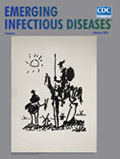
Commemorating Misadventures, Celebrating Collaborations [PDF - 2.19 MB - 2 pages]
| EID | Breedlove B. Commemorating Misadventures, Celebrating Collaborations. Emerg Infect Dis. 2018;24(2):407-408. https://doi.org/10.3201/eid2402.ac2402 |
|---|---|
| AMA | Breedlove B. Commemorating Misadventures, Celebrating Collaborations. Emerging Infectious Diseases. 2018;24(2):407-408. doi:10.3201/eid2402.ac2402. |
| APA | Breedlove, B. (2018). Commemorating Misadventures, Celebrating Collaborations. Emerging Infectious Diseases, 24(2), 407-408. https://doi.org/10.3201/eid2402.ac2402. |


