Perspective
Ferrets as Models for Influenza Virus Transmission Studies and Pandemic Risk Assessments [PDF - 424 KB - 7 pages]
The ferret transmission model is extensively used to assess the pandemic potential of emerging influenza viruses, yet experimental conditions and reported results vary among laboratories. Such variation can be a critical consideration when contextualizing results from independent risk-assessment studies of novel and emerging influenza viruses. To streamline interpretation of data generated in different laboratories, we provide a consensus on experimental parameters that define risk-assessment experiments of influenza virus transmissibility, including disclosure of variables known or suspected to contribute to experimental variability in this model, and advocate adoption of more standardized practices. We also discuss current limitations of the ferret transmission model and highlight continued refinements and advances to this model ongoing in laboratories. Understanding, disclosing, and standardizing the critical parameters of ferret transmission studies will improve the comparability and reproducibility of pandemic influenza risk assessment and increase the statistical power and, perhaps, accuracy of this model.
| EID | Belser JA, Barclay W, Barr I, Fouchier R, Matsuyama R, Nishiura H, et al. Ferrets as Models for Influenza Virus Transmission Studies and Pandemic Risk Assessments. Emerg Infect Dis. 2018;24(6):965-971. https://doi.org/10.3201/eid2406.172114 |
|---|---|
| AMA | Belser JA, Barclay W, Barr I, et al. Ferrets as Models for Influenza Virus Transmission Studies and Pandemic Risk Assessments. Emerging Infectious Diseases. 2018;24(6):965-971. doi:10.3201/eid2406.172114. |
| APA | Belser, J. A., Barclay, W., Barr, I., Fouchier, R., Matsuyama, R., Nishiura, H....Yen, H. (2018). Ferrets as Models for Influenza Virus Transmission Studies and Pandemic Risk Assessments. Emerging Infectious Diseases, 24(6), 965-971. https://doi.org/10.3201/eid2406.172114. |
Synopses
Absence of Nosocomial Transmission of Imported Lassa Fever during Use of Standard Barrier Nursing Methods [PDF - 1.22 MB - 6 pages]
Nosocomial transmission of Lassa virus (LASV) is reported to be low when care for the index patient includes proper barrier nursing methods. We investigated whether asymptomatic LASV infection occurred in healthcare workers who used standard barrier nursing methods during the first 15 days of caring for a patient with Lassa fever in Sweden. Of 76 persons who were defined as having been potentially exposed to LASV, 53 provided blood samples for detection of LASV IgG. These persons also responded to a detailed questionnaire to evaluate exposure to different body fluids from the index patient. LASV-specific IgG was not detected in any of the 53 persons. Five of 53 persons had not been using proper barrier nursing methods. Our results strengthen the argument for a low risk of secondary transmission of LASV in humans when standard barrier nursing methods are used and the patient has only mild symptoms.
| EID | Grahn A, Bråve A, Tolfvenstam T, Studahl M. Absence of Nosocomial Transmission of Imported Lassa Fever during Use of Standard Barrier Nursing Methods. Emerg Infect Dis. 2018;24(6):972-977. https://doi.org/10.3201/eid2406.172097 |
|---|---|
| AMA | Grahn A, Bråve A, Tolfvenstam T, et al. Absence of Nosocomial Transmission of Imported Lassa Fever during Use of Standard Barrier Nursing Methods. Emerging Infectious Diseases. 2018;24(6):972-977. doi:10.3201/eid2406.172097. |
| APA | Grahn, A., Bråve, A., Tolfvenstam, T., & Studahl, M. (2018). Absence of Nosocomial Transmission of Imported Lassa Fever during Use of Standard Barrier Nursing Methods. Emerging Infectious Diseases, 24(6), 972-977. https://doi.org/10.3201/eid2406.172097. |
Research
Limbic encephalitis is commonly regarded as an autoimmune-mediated disease. However, after the recent detection of zoonotic variegated squirrel bornavirus 1 in a Prevost’s squirrel (Callosciurus prevostii) in a zoo in northern Germany, we retrospectively investigated a fatal case in an autoantibody-seronegative animal caretaker who had worked at that zoo. The virus had been discovered in 2015 as the cause of a cluster of cases of fatal encephalitis among breeders of variegated squirrels (Sciurus variegatoides) in eastern Germany. Molecular assays and immunohistochemistry detected a limbic distribution of the virus in brain tissue of the animal caretaker. Phylogenetic analyses demonstrated a spillover infection from the Prevost’s squirrel. Antibodies against bornaviruses were detected in the patient’s cerebrospinal fluid by immunofluorescence and newly developed ELISAs and immunoblot. The putative antigenic epitope was identified on the viral nucleoprotein. Other zoo workers were not infected; however, avoidance of direct contact with exotic squirrels and screening of squirrels are recommended.
| EID | Tappe D, Schlottau K, Cadar D, Hoffmann B, Balke L, Bewig B, et al. Occupation-Associated Fatal Limbic Encephalitis Caused by Variegated Squirrel Bornavirus 1, Germany, 2013. Emerg Infect Dis. 2018;24(6):978-987. https://doi.org/10.3201/eid2406.172027 |
|---|---|
| AMA | Tappe D, Schlottau K, Cadar D, et al. Occupation-Associated Fatal Limbic Encephalitis Caused by Variegated Squirrel Bornavirus 1, Germany, 2013. Emerging Infectious Diseases. 2018;24(6):978-987. doi:10.3201/eid2406.172027. |
| APA | Tappe, D., Schlottau, K., Cadar, D., Hoffmann, B., Balke, L., Bewig, B....Beer, M. (2018). Occupation-Associated Fatal Limbic Encephalitis Caused by Variegated Squirrel Bornavirus 1, Germany, 2013. Emerging Infectious Diseases, 24(6), 978-987. https://doi.org/10.3201/eid2406.172027. |
Genomic Sequencing of Bordetella pertussis for Epidemiology and Global Surveillance of Whooping Cough [PDF - 1.37 MB - 7 pages]
Bordetella pertussis causes whooping cough, a highly contagious respiratory disease that is reemerging in many world regions. The spread of antigen-deficient strains may threaten acellular vaccine efficacy. Dynamics of strain transmission are poorly defined because of shortcomings in current strain genotyping methods. Our objective was to develop a whole-genome genotyping strategy with sufficient resolution for local epidemiologic questions and sufficient reproducibility to enable international comparisons of clinical isolates. We defined a core genome multilocus sequence typing scheme comprising 2,038 loci and demonstrated its congruence with whole-genome single-nucleotide polymorphism variation. Most cases of intrafamilial groups of isolates or of multiple isolates recovered from the same patient were distinguished from temporally and geographically cocirculating isolates. However, epidemiologically unrelated isolates were sometimes nearly undistinguishable. We set up a publicly accessible core genome multilocus sequence typing database to enable global comparisons of B. pertussis isolates, opening the way for internationally coordinated surveillance.
| EID | Bouchez V, Guglielmini J, Dazas M, Landier A, Toubiana J, Guillot S, et al. Genomic Sequencing of Bordetella pertussis for Epidemiology and Global Surveillance of Whooping Cough. Emerg Infect Dis. 2018;24(6):988-994. https://doi.org/10.3201/eid2406.171464 |
|---|---|
| AMA | Bouchez V, Guglielmini J, Dazas M, et al. Genomic Sequencing of Bordetella pertussis for Epidemiology and Global Surveillance of Whooping Cough. Emerging Infectious Diseases. 2018;24(6):988-994. doi:10.3201/eid2406.171464. |
| APA | Bouchez, V., Guglielmini, J., Dazas, M., Landier, A., Toubiana, J., Guillot, S....Brisse, S. (2018). Genomic Sequencing of Bordetella pertussis for Epidemiology and Global Surveillance of Whooping Cough. Emerging Infectious Diseases, 24(6), 988-994. https://doi.org/10.3201/eid2406.171464. |
Use of Bead-Based Serologic Assay to Evaluate Chikungunya Virus Epidemic, Haiti [PDF - 2.31 MB - 7 pages]
The index case of chikungunya virus (CHIKV) in Haiti was reported during early 2014; the vector, the pervasive Aedes aegypti mosquito, promoted rapid spread throughout the country. During December 2014–February 2015, we collected blood samples from 4,438 persons at 154 sites (62 urban, 92 rural) throughout Haiti and measured CHIKV IgG by using a multiplex bead assay. Overall CHIKV seroprevalence was 57.9%; differences between rural (mean 44.9%) and urban (mean 78.4%) areas were pronounced. Logistic modeling identified the urban environment as a strong predictor of CHIKV exposure (adjusted odds ratio 3.34, 95% CI 2.38–4.69), and geographic elevation provided a strong negative correlation. We observed no correlation between age and antibody positivity or titer. Our findings demonstrated through serologic testing the recent and rapid dissemination of the arbovirus throughout the country. These results show the utility of serologic data to conduct epidemiologic studies of quickly spreading mosquitoborne arboviruses.
| EID | Rogier EW, Moss DM, Mace KE, Chang M, Jean SE, Bullard SM, et al. Use of Bead-Based Serologic Assay to Evaluate Chikungunya Virus Epidemic, Haiti. Emerg Infect Dis. 2018;24(6):995-1001. https://doi.org/10.3201/eid2406.171447 |
|---|---|
| AMA | Rogier EW, Moss DM, Mace KE, et al. Use of Bead-Based Serologic Assay to Evaluate Chikungunya Virus Epidemic, Haiti. Emerging Infectious Diseases. 2018;24(6):995-1001. doi:10.3201/eid2406.171447. |
| APA | Rogier, E. W., Moss, D. M., Mace, K. E., Chang, M., Jean, S. E., Bullard, S. M....Udhayakumar, V. (2018). Use of Bead-Based Serologic Assay to Evaluate Chikungunya Virus Epidemic, Haiti. Emerging Infectious Diseases, 24(6), 995-1001. https://doi.org/10.3201/eid2406.171447. |
Widespread Treponema pallidum Infection in Nonhuman Primates, Tanzania [PDF - 2.03 MB - 8 pages]
We investigated Treponema pallidum infection in 8 nonhuman primate species (289 animals) in Tanzania during 2015–2017. We used a serologic treponemal test to detect antibodies against the bacterium. Infection was further confirmed from tissue samples of skin-ulcerated animals by 3 independent PCRs (polA, tp47, and TP_0619). Our findings indicate that T. pallidum infection is geographically widespread in Tanzania and occurs in several species (olive baboons, yellow baboons, vervet monkeys, and blue monkeys). We found the bacterium at 11 of 14 investigated geographic locations. Anogenital ulceration was the most common clinical manifestation; orofacial lesions also were observed. Molecular data show that nonhuman primates in Tanzania are most likely infected with T. pallidum subsp. pertenue–like strains, which could have implications for human yaws eradication.
| EID | Chuma IS, Batamuzi EK, Collins D, Fyumagwa RD, Hallmaier-Wacker LK, Kazwala RR, et al. Widespread Treponema pallidum Infection in Nonhuman Primates, Tanzania. Emerg Infect Dis. 2018;24(6):1002-1009. https://doi.org/10.3201/eid2406.180037 |
|---|---|
| AMA | Chuma IS, Batamuzi EK, Collins D, et al. Widespread Treponema pallidum Infection in Nonhuman Primates, Tanzania. Emerging Infectious Diseases. 2018;24(6):1002-1009. doi:10.3201/eid2406.180037. |
| APA | Chuma, I. S., Batamuzi, E. K., Collins, D., Fyumagwa, R. D., Hallmaier-Wacker, L. K., Kazwala, R. R....Knauf, S. (2018). Widespread Treponema pallidum Infection in Nonhuman Primates, Tanzania. Emerging Infectious Diseases, 24(6), 1002-1009. https://doi.org/10.3201/eid2406.180037. |
Genomic Epidemiology of Global Carbapenemase-Producing Enterobacter spp., 2008–2014 [PDF - 5.26 MB - 10 pages]
We performed whole-genome sequencing on 170 clinical carbapenemase-producing Enterobacter spp. isolates collected globally during 2008–2014. The most common carbapenemase was VIM, followed by New Delhi metallo-β-lactamase (NDM), Klebsiella pneumoniae carbapenemase, oxacillin 48, and IMP. The isolates were of predominantly 2 species (E. xiangfangensis and E. hormaechei subsp. steigerwaltii) and 4 global clones (sequence type [ST] 114, ST93, ST90, and ST78) with different clades within ST114 and ST90. Particular genetic structures surrounding carbapenemase genes were circulating locally in various institutions within the same or between different STs in Greece, Guatemala, Italy, Spain, Serbia, and Vietnam. We found a common NDM genetic structure (NDM-GE-U.S.), previously described on pNDM-U.S. from Klebsiella pneumoniae ATCC BAA-214, in 14 different clones obtained from 6 countries spanning 4 continents. Our study highlights the importance of surveillance programs using whole-genome sequencing in providing insight into the molecular epidemiology of carbapenemase-producing Enterobacter spp.
| EID | Peirano G, Matsumura Y, Adams MD, Bradford P, Motyl M, Chen L, et al. Genomic Epidemiology of Global Carbapenemase-Producing Enterobacter spp., 2008–2014. Emerg Infect Dis. 2018;24(6):1010-1019. https://doi.org/10.3201/eid2406.171648 |
|---|---|
| AMA | Peirano G, Matsumura Y, Adams MD, et al. Genomic Epidemiology of Global Carbapenemase-Producing Enterobacter spp., 2008–2014. Emerging Infectious Diseases. 2018;24(6):1010-1019. doi:10.3201/eid2406.171648. |
| APA | Peirano, G., Matsumura, Y., Adams, M. D., Bradford, P., Motyl, M., Chen, L....Pitout, J. (2018). Genomic Epidemiology of Global Carbapenemase-Producing Enterobacter spp., 2008–2014. Emerging Infectious Diseases, 24(6), 1010-1019. https://doi.org/10.3201/eid2406.171648. |
Influenza D Virus Infection in Feral Swine Populations, United States [PDF - 3.18 MB - 9 pages]
Influenza D virus (IDV) has been identified in domestic cattle, swine, camelid, and small ruminant populations across North America, Europe, Asia, South America, and Africa. Our study investigated seroprevalence and transmissibility of IDV in feral swine. During 2012–2013, we evaluated feral swine populations in 4 US states; of 256 swine tested, 57 (19.1%) were IDV seropositive. Among 96 archived influenza A virus–seropositive feral swine samples collected from 16 US states during 2010–2013, 41 (42.7%) were IDV seropositive. Infection studies demonstrated that IDV-inoculated feral swine shed virus 3–5 days postinoculation and seroconverted at 21 days postinoculation; 50% of in-contact naive feral swine shed virus, seroconverted, or both. Immunohistochemical staining showed viral antigen within epithelial cells of the respiratory tract, including trachea, soft palate, and lungs. Our findings suggest that feral swine might serve an important role in the ecology of IDV.
| EID | Ferguson L, Luo K, Olivier AK, Cunningham FL, Blackmon S, Hanson-Dorr K, et al. Influenza D Virus Infection in Feral Swine Populations, United States. Emerg Infect Dis. 2018;24(6):1020-1028. https://doi.org/10.3201/eid2406.172102 |
|---|---|
| AMA | Ferguson L, Luo K, Olivier AK, et al. Influenza D Virus Infection in Feral Swine Populations, United States. Emerging Infectious Diseases. 2018;24(6):1020-1028. doi:10.3201/eid2406.172102. |
| APA | Ferguson, L., Luo, K., Olivier, A. K., Cunningham, F. L., Blackmon, S., Hanson-Dorr, K....Wan, X. (2018). Influenza D Virus Infection in Feral Swine Populations, United States. Emerging Infectious Diseases, 24(6), 1020-1028. https://doi.org/10.3201/eid2406.172102. |
Prion Disease in Dromedary Camels, Algeria [PDF - 3.73 MB - 8 pages]
Prions cause fatal and transmissible neurodegenerative diseases, including Creutzfeldt-Jakob disease in humans, scrapie in small ruminants, and bovine spongiform encephalopathy (BSE). After the BSE epidemic, and the associated human infections, began in 1996 in the United Kingdom, general concerns have been raised about animal prions. We detected a prion disease in dromedary camels (Camelus dromedarius) in Algeria. Symptoms suggesting prion disease occurred in 3.1% of dromedaries brought for slaughter to the Ouargla abattoir in 2015–2016. We confirmed diagnosis by detecting pathognomonic neurodegeneration and disease-specific prion protein (PrPSc) in brain tissues from 3 symptomatic animals. Prion detection in lymphoid tissues is suggestive of the infectious nature of the disease. PrPSc biochemical characterization showed differences with BSE and scrapie. Our identification of this prion disease in a geographically widespread livestock species requires urgent enforcement of surveillance and assessment of the potential risks to human and animal health.
| EID | Babelhadj B, Di Bari M, Pirisinu L, Chiappini B, Gaouar S, Riccardi G, et al. Prion Disease in Dromedary Camels, Algeria. Emerg Infect Dis. 2018;24(6):1029-1036. https://doi.org/10.3201/eid2406.172007 |
|---|---|
| AMA | Babelhadj B, Di Bari M, Pirisinu L, et al. Prion Disease in Dromedary Camels, Algeria. Emerging Infectious Diseases. 2018;24(6):1029-1036. doi:10.3201/eid2406.172007. |
| APA | Babelhadj, B., Di Bari, M., Pirisinu, L., Chiappini, B., Gaouar, S., Riccardi, G....Vaccari, G. (2018). Prion Disease in Dromedary Camels, Algeria. Emerging Infectious Diseases, 24(6), 1029-1036. https://doi.org/10.3201/eid2406.172007. |
Frequent Implication of Multistress-Tolerant Campylobacter jejuni in Human Infections [PDF - 2.50 MB - 8 pages]
Campylobacter jejuni, a major cause of bacterial foodborne illnesses, is considered highly susceptible to environmental stresses. In this study, we extensively investigated the stress tolerance of 121 clinical strains of C. jejuni against 5 stress conditions (aerobic stress, disinfectant exposure, freeze-thaw, heat treatment, and osmotic stress) that this pathogenic bacterium might encounter during foodborne transmission to humans. In contrast to our current perception about high stress sensitivity of C. jejuni, a number of clinical strains of C. jejuni were highly tolerant to multiple stresses. We performed population genetics analysis by using comparative genomic fingerprinting and showed that multistress-tolerant strains of C. jejuni constituted distinct clades. The comparative genomic fingerprinting subtypes belonging to multistress-tolerant clades were more frequently implicated in human infections than those in stress-sensitive clades. We identified unique stress-tolerant C. jejuni clones and showed the role of stress tolerance in human campylobacteriosis.
| EID | Oh E, Chui L, Bae J, Li V, Ma A, Mutschall SK, et al. Frequent Implication of Multistress-Tolerant Campylobacter jejuni in Human Infections. Emerg Infect Dis. 2018;24(6):1037-1044. https://doi.org/10.3201/eid2406.171587 |
|---|---|
| AMA | Oh E, Chui L, Bae J, et al. Frequent Implication of Multistress-Tolerant Campylobacter jejuni in Human Infections. Emerging Infectious Diseases. 2018;24(6):1037-1044. doi:10.3201/eid2406.171587. |
| APA | Oh, E., Chui, L., Bae, J., Li, V., Ma, A., Mutschall, S. K....Jeon, B. (2018). Frequent Implication of Multistress-Tolerant Campylobacter jejuni in Human Infections. Emerging Infectious Diseases, 24(6), 1037-1044. https://doi.org/10.3201/eid2406.171587. |
We conducted a multicenter, retrospective cohort study of hospitalized patients with serologically proven nephropathia epidemica (NE) living in Ardennes Department, France, during 2000–2014 to develop a bioclinical test predictive of severe disease. Among 205 patients, 45 (22.0%) had severe NE. We found the following factors predictive of severe NE: nephrotoxic drug exposure (p = 0.005, point value 10); visual disorders (p = 0.02, point value 8); microscopic or macroscopic hematuria (p = 0.04, point value 7); leukocyte count >10 × 109 cells/L (p = 0.01, point value 9); and thrombocytopenia <90 × 109/L (p = 0.003, point value 11). When point values for each factor were summed, we found a score of <10 identified low-risk patients (3.3% had severe disease), and a score >20 identified high-risk patients (45.3% had severe disease). If validated in future studies, this test could be used to stratify patients by severity in research studies and in clinical practice.
| EID | Hentzien M, Mestrallet S, Halin P, Pannet L, Lebrun D, Dramé M, et al. Bioclinical Test to Predict Nephropathia Epidemica Severity at Hospital Admission. Emerg Infect Dis. 2018;24(6):1045-1054. https://doi.org/10.3201/eid2406.172160 |
|---|---|
| AMA | Hentzien M, Mestrallet S, Halin P, et al. Bioclinical Test to Predict Nephropathia Epidemica Severity at Hospital Admission. Emerging Infectious Diseases. 2018;24(6):1045-1054. doi:10.3201/eid2406.172160. |
| APA | Hentzien, M., Mestrallet, S., Halin, P., Pannet, L., Lebrun, D., Dramé, M....Servettaz, A. (2018). Bioclinical Test to Predict Nephropathia Epidemica Severity at Hospital Admission. Emerging Infectious Diseases, 24(6), 1045-1054. https://doi.org/10.3201/eid2406.172160. |
Hepatitis E in Long-Term Travelers from the Netherlands to Subtropical and Tropical Countries, 2008–2011 [PDF - 765 KB - 6 pages]
Hepatitis E virus (HEV) is a common cause of acute viral hepatitis. Virus genotypes 1 and 2 infect humans in developing countries by the fecal–oral route. To assess attack rates and disease incidence for travelers, we prospectively studied 604 long-term travelers to subtropical and tropical countries. Participants donated blood samples pretravel and posttravel and kept a diary. A total of 89/604 (15%) pretravel samples were positive for HEV IgG by ELISA, suggesting previous HEV infection. Seroconversion for HEV was found for 19/515 travelers (attack rate 3.7%, incidence 1.8 cases/1,000 person-weeks). We believe there is a substantial risk for acquiring HEV infection among long-term travelers. Although HEV infection does not seem to be a major problem in this healthy cohort, hygienic measures should be stressed in all pretravel health advice, particularly for pregnant women and immunocompromised travelers who are at risk for severe disease.
| EID | Elfrink F, Overbosch FW, Schinkel J, Koen G, Sonder G. Hepatitis E in Long-Term Travelers from the Netherlands to Subtropical and Tropical Countries, 2008–2011. Emerg Infect Dis. 2018;24(6):1055-1060. https://doi.org/10.3201/eid2406.171513 |
|---|---|
| AMA | Elfrink F, Overbosch FW, Schinkel J, et al. Hepatitis E in Long-Term Travelers from the Netherlands to Subtropical and Tropical Countries, 2008–2011. Emerging Infectious Diseases. 2018;24(6):1055-1060. doi:10.3201/eid2406.171513. |
| APA | Elfrink, F., Overbosch, F. W., Schinkel, J., Koen, G., & Sonder, G. (2018). Hepatitis E in Long-Term Travelers from the Netherlands to Subtropical and Tropical Countries, 2008–2011. Emerging Infectious Diseases, 24(6), 1055-1060. https://doi.org/10.3201/eid2406.171513. |
Novel Parvovirus Related to Primate Bufaviruses in Dogs [PDF - 1.21 MB - 8 pages]
A novel protoparvovirus species, related genetically to human bufaviruses, was identified in dogs with respiratory signs. The canine bufavirus was distantly related to the well-known canine protoparvovirus, canine parvovirus type 2, sharing low amino acid identities in the nonstructural protein 1 (40.6%) and in the capsid protein 1 (33.4%). By screening collections of fecal, nasal, and oropharyngeal samples obtained from juvenile dogs (<1 year of age), canine bufavirus DNA appeared as a common component of canine virome. The virus was common in the stool samples of dogs with or without enteric disease and in the nasal and oropharyngeal swab samples of dogs with respiratory signs. However, the virus was not detected in nasal and oropharyngeal swab samples from animals without clinical signs.
| EID | Martella V, Lanave G, Mihalov-Kovács E, Marton S, Varga-Kugler R, Kaszab E, et al. Novel Parvovirus Related to Primate Bufaviruses in Dogs. Emerg Infect Dis. 2018;24(6):1061-1068. https://doi.org/10.3201/eid2406.171965 |
|---|---|
| AMA | Martella V, Lanave G, Mihalov-Kovács E, et al. Novel Parvovirus Related to Primate Bufaviruses in Dogs. Emerging Infectious Diseases. 2018;24(6):1061-1068. doi:10.3201/eid2406.171965. |
| APA | Martella, V., Lanave, G., Mihalov-Kovács, E., Marton, S., Varga-Kugler, R., Kaszab, E....Bányai, K. (2018). Novel Parvovirus Related to Primate Bufaviruses in Dogs. Emerging Infectious Diseases, 24(6), 1061-1068. https://doi.org/10.3201/eid2406.171965. |
Dispatches
Novel Poxvirus in Proliferative Lesions of Wild Rodents in East Central Texas, USA [PDF - 2.30 MB - 4 pages]
Northern pygmy mice from 2 localities in East Central Texas, USA, had proliferative epidermal lesions on the tail and feet. Electron microscopy of lesion tissue revealed poxvirus. Phylogenetic analyses indicated the virus differed 35% from its closest relatives, the Chordopoxvirinae. Future research is needed to determine whether this virus could affect human health.
| EID | Hodo CL, Mauldin MR, Light JE, Wilkins K, Tang S, Nakazawa Y, et al. Novel Poxvirus in Proliferative Lesions of Wild Rodents in East Central Texas, USA. Emerg Infect Dis. 2018;24(6):1069-1072. https://doi.org/10.3201/eid2406.172057 |
|---|---|
| AMA | Hodo CL, Mauldin MR, Light JE, et al. Novel Poxvirus in Proliferative Lesions of Wild Rodents in East Central Texas, USA. Emerging Infectious Diseases. 2018;24(6):1069-1072. doi:10.3201/eid2406.172057. |
| APA | Hodo, C. L., Mauldin, M. R., Light, J. E., Wilkins, K., Tang, S., Nakazawa, Y....Hamer, S. A. (2018). Novel Poxvirus in Proliferative Lesions of Wild Rodents in East Central Texas, USA. Emerging Infectious Diseases, 24(6), 1069-1072. https://doi.org/10.3201/eid2406.172057. |
Foot-and-Mouth Disease in the Middle East Caused by an A/ASIA/G-VII Virus Lineage, 2015–2016 [PDF - 3.50 MB - 6 pages]
Phylogenetic analyses of foot-and-mouth disease type A viruses in the Middle East during 2015–2016 identified viruses belonging to the A/ASIA/G-VII lineage, which originated in the Indian subcontinent. Changes in a critical antigenic site within capsid viral protein 1 suggest possible evolutionary pressure caused by an intensive vaccination program.
| EID | Bachanek-Bankowska K, Di Nardo A, Wadsworth J, Henry E, Parlak Ü, Timina A, et al. Foot-and-Mouth Disease in the Middle East Caused by an A/ASIA/G-VII Virus Lineage, 2015–2016. Emerg Infect Dis. 2018;24(6):1073-1078. https://doi.org/10.3201/eid2406.170715 |
|---|---|
| AMA | Bachanek-Bankowska K, Di Nardo A, Wadsworth J, et al. Foot-and-Mouth Disease in the Middle East Caused by an A/ASIA/G-VII Virus Lineage, 2015–2016. Emerging Infectious Diseases. 2018;24(6):1073-1078. doi:10.3201/eid2406.170715. |
| APA | Bachanek-Bankowska, K., Di Nardo, A., Wadsworth, J., Henry, E., Parlak, Ü., Timina, A....Knowles, N. J. (2018). Foot-and-Mouth Disease in the Middle East Caused by an A/ASIA/G-VII Virus Lineage, 2015–2016. Emerging Infectious Diseases, 24(6), 1073-1078. https://doi.org/10.3201/eid2406.170715. |
Novel Salmonella enterica Serovar Typhimurium Genotype Levels as Herald of Seasonal Salmonellosis Epidemics [PDF - 1.83 MB - 4 pages]
We examined the population dynamics of Salmonella enterica serovar Typhimurium during seasonal salmonellosis epidemics in New South Wales, Australia, during 2009–2016. Of 15,626 isolates, 5%–20% consisted of novel genotypes. Seasons with salmonellosis epidemics were associated with a reduction in novel genotypes in the preceding winter and spring.
| EID | Sotomayor C, Wang Q, Arnott A, Howard P, Hope K, Lan R, et al. Novel Salmonella enterica Serovar Typhimurium Genotype Levels as Herald of Seasonal Salmonellosis Epidemics. Emerg Infect Dis. 2018;24(6):1079-1082. https://doi.org/10.3201/eid2406.171096 |
|---|---|
| AMA | Sotomayor C, Wang Q, Arnott A, et al. Novel Salmonella enterica Serovar Typhimurium Genotype Levels as Herald of Seasonal Salmonellosis Epidemics. Emerging Infectious Diseases. 2018;24(6):1079-1082. doi:10.3201/eid2406.171096. |
| APA | Sotomayor, C., Wang, Q., Arnott, A., Howard, P., Hope, K., Lan, R....Sintchenko, V. (2018). Novel Salmonella enterica Serovar Typhimurium Genotype Levels as Herald of Seasonal Salmonellosis Epidemics. Emerging Infectious Diseases, 24(6), 1079-1082. https://doi.org/10.3201/eid2406.171096. |
Urban Wild Boars and Risk for Zoonotic Streptococcus suis, Spain [PDF - 1.47 MB - 4 pages]
Urban wild boars (Sus scrofa) from Barcelona, Spain, harbor great diversity of Streptococcus suis strains, including strains with the cps2 gene and with the same molecular profile as local human cases. The increasing trend of potential effective contacts for S. suis transmission is of public health concern.
| EID | Fernández-Aguilar X, Gottschalk M, Aragon V, Càmara J, Ardanuy C, Velarde R, et al. Urban Wild Boars and Risk for Zoonotic Streptococcus suis, Spain. Emerg Infect Dis. 2018;24(6):1083-1086. https://doi.org/10.3201/eid2406.171271 |
|---|---|
| AMA | Fernández-Aguilar X, Gottschalk M, Aragon V, et al. Urban Wild Boars and Risk for Zoonotic Streptococcus suis, Spain. Emerging Infectious Diseases. 2018;24(6):1083-1086. doi:10.3201/eid2406.171271. |
| APA | Fernández-Aguilar, X., Gottschalk, M., Aragon, V., Càmara, J., Ardanuy, C., Velarde, R....Cabezón, O. (2018). Urban Wild Boars and Risk for Zoonotic Streptococcus suis, Spain. Emerging Infectious Diseases, 24(6), 1083-1086. https://doi.org/10.3201/eid2406.171271. |
Human Endophthalmitis Caused By Pseudorabies Virus Infection, China, 2017 [PDF - 1.43 MB - 4 pages]
We report human endophthalmitis caused by pseudorabies virus infection after exposure to sewage on a hog farm in China. High-throughput sequencing and real-time PCR of vitreous humor showed pseudorabies virus sequences. This case showed that pseudorabies virus might infect humans after direct contact with contaminants.
| EID | Ai J, Weng S, Cheng Q, Cui P, Li Y, Wu H, et al. Human Endophthalmitis Caused By Pseudorabies Virus Infection, China, 2017. Emerg Infect Dis. 2018;24(6):1087-1090. https://doi.org/10.3201/eid2406.171612 |
|---|---|
| AMA | Ai J, Weng S, Cheng Q, et al. Human Endophthalmitis Caused By Pseudorabies Virus Infection, China, 2017. Emerging Infectious Diseases. 2018;24(6):1087-1090. doi:10.3201/eid2406.171612. |
| APA | Ai, J., Weng, S., Cheng, Q., Cui, P., Li, Y., Wu, H....Zhang, W. (2018). Human Endophthalmitis Caused By Pseudorabies Virus Infection, China, 2017. Emerging Infectious Diseases, 24(6), 1087-1090. https://doi.org/10.3201/eid2406.171612. |
Pulmonary Infections with Nontuberculous Mycobacteria, Catalonia, Spain, 1994–2014 [PDF - 1.20 MB - 4 pages]
In Spain, systematic reporting of pulmonary infections with nontuberculous mycobacteria is not mandatory. Therefore, to determine trends, we retrospectively identified cases for January 1994–December 2014 in Catalonia. Over the 21 years, prevalence increased and was associated with being male. Mycobacterium avium complex and M. abscessus prevalence increased; M. kansasii prevalence decreased.
| EID | Santin M, Barrabeig I, Malchair P, Gonzalez-Luquero L, Benitez MA, Sabria J, et al. Pulmonary Infections with Nontuberculous Mycobacteria, Catalonia, Spain, 1994–2014. Emerg Infect Dis. 2018;24(6):1091-1094. https://doi.org/10.3201/eid2406.172095 |
|---|---|
| AMA | Santin M, Barrabeig I, Malchair P, et al. Pulmonary Infections with Nontuberculous Mycobacteria, Catalonia, Spain, 1994–2014. Emerging Infectious Diseases. 2018;24(6):1091-1094. doi:10.3201/eid2406.172095. |
| APA | Santin, M., Barrabeig, I., Malchair, P., Gonzalez-Luquero, L., Benitez, M. A., Sabria, J....Alcaide, F. (2018). Pulmonary Infections with Nontuberculous Mycobacteria, Catalonia, Spain, 1994–2014. Emerging Infectious Diseases, 24(6), 1091-1094. https://doi.org/10.3201/eid2406.172095. |
Westward Spread of Highly Pathogenic Avian Influenza A(H7N9) Virus among Humans, China [PDF - 1.53 MB - 4 pages]
We report infection of humans with highly pathogenic avian influenza A(H7N9) virus in Shaanxi, China, in May 2017. We obtained complete genomes for samples from 5 patients and from live poultry markets or farms in 4 cities. Results indicate that H7N9 is spreading westward from southern and eastern China.
| EID | Yang Q, Shi W, Zhang L, Xu Y, Xu J, Li S, et al. Westward Spread of Highly Pathogenic Avian Influenza A(H7N9) Virus among Humans, China. Emerg Infect Dis. 2018;24(6):1095-1098. https://doi.org/10.3201/eid2406.171135 |
|---|---|
| AMA | Yang Q, Shi W, Zhang L, et al. Westward Spread of Highly Pathogenic Avian Influenza A(H7N9) Virus among Humans, China. Emerging Infectious Diseases. 2018;24(6):1095-1098. doi:10.3201/eid2406.171135. |
| APA | Yang, Q., Shi, W., Zhang, L., Xu, Y., Xu, J., Li, S....Tian, H. (2018). Westward Spread of Highly Pathogenic Avian Influenza A(H7N9) Virus among Humans, China. Emerging Infectious Diseases, 24(6), 1095-1098. https://doi.org/10.3201/eid2406.171135. |
Importation of Human Seoul Virus Infection to Germany from Indonesia [PDF - 2.40 MB - 4 pages]
Seoul hantavirus–associated hemorrhagic fever with renal syndrome cases are rare outside Asia and have not yet been found in Germany. We report clinical and molecular evidence for a Seoul virus infection in a patient in Germany. The infection was most likely acquired during a stay in Sulawesi, Indonesia.
| EID | Hofmann J, Weiss S, Kuhns M, Zinke A, Heinsberger H, Kruger DH. Importation of Human Seoul Virus Infection to Germany from Indonesia. Emerg Infect Dis. 2018;24(6):1099-1102. https://doi.org/10.3201/eid2406.172044 |
|---|---|
| AMA | Hofmann J, Weiss S, Kuhns M, et al. Importation of Human Seoul Virus Infection to Germany from Indonesia. Emerging Infectious Diseases. 2018;24(6):1099-1102. doi:10.3201/eid2406.172044. |
| APA | Hofmann, J., Weiss, S., Kuhns, M., Zinke, A., Heinsberger, H., & Kruger, D. H. (2018). Importation of Human Seoul Virus Infection to Germany from Indonesia. Emerging Infectious Diseases, 24(6), 1099-1102. https://doi.org/10.3201/eid2406.172044. |
Rickettsia parkeri in Dermacentor parumapertus Ticks, Mexico [PDF - 1.47 MB - 4 pages]
During a study to identify zoonotic pathogens in northwestern Mexico, we detected the presence of a rickettsial agent in Dermacentor parumapertus ticks from black-tailed jackrabbits (Lepus californicus). Comparison of 4 gene sequences (gltA, htrA, ompA, and ompB) of this agent showed 99%–100% identity with sequences of Rickettsia parkeri.
| EID | Sánchez-Montes S, López-Pérez AM, Guzmán-Cornejo C, Colunga-Salas P, Becker I, Delgado-de la Mora J, et al. Rickettsia parkeri in Dermacentor parumapertus Ticks, Mexico. Emerg Infect Dis. 2018;24(6):1108-1111. https://doi.org/10.3201/eid2406.180058 |
|---|---|
| AMA | Sánchez-Montes S, López-Pérez AM, Guzmán-Cornejo C, et al. Rickettsia parkeri in Dermacentor parumapertus Ticks, Mexico. Emerging Infectious Diseases. 2018;24(6):1108-1111. doi:10.3201/eid2406.180058. |
| APA | Sánchez-Montes, S., López-Pérez, A. M., Guzmán-Cornejo, C., Colunga-Salas, P., Becker, I., Delgado-de la Mora, J....Suzán, G. (2018). Rickettsia parkeri in Dermacentor parumapertus Ticks, Mexico. Emerging Infectious Diseases, 24(6), 1108-1111. https://doi.org/10.3201/eid2406.180058. |
Novel Focus of Sin Nombre Virus in Peromyscus eremicus Mice, Death Valley National Park, California, USA [PDF - 2.32 MB - 4 pages]
The deer mouse (Peromyscus maniculatus) is the primary reservoir for Sin Nombre virus (SNV) in the western United States. Rodent surveillance for hantavirus in Death Valley National Park, California, USA, revealed cactus mice (P. eremicus) as a possible focal reservoir for SNV in this location. We identified SNV antibodies in 40% of cactus mice sampled.
| EID | Burns JE, Metzger ME, Messenger S, Fritz CL, Vilcins IE, Enge B, et al. Novel Focus of Sin Nombre Virus in Peromyscus eremicus Mice, Death Valley National Park, California, USA. Emerg Infect Dis. 2018;24(6):1112-1115. https://doi.org/10.3201/eid2406.180089 |
|---|---|
| AMA | Burns JE, Metzger ME, Messenger S, et al. Novel Focus of Sin Nombre Virus in Peromyscus eremicus Mice, Death Valley National Park, California, USA. Emerging Infectious Diseases. 2018;24(6):1112-1115. doi:10.3201/eid2406.180089. |
| APA | Burns, J. E., Metzger, M. E., Messenger, S., Fritz, C. L., Vilcins, I. E., Enge, B....Hu, R. (2018). Novel Focus of Sin Nombre Virus in Peromyscus eremicus Mice, Death Valley National Park, California, USA. Emerging Infectious Diseases, 24(6), 1112-1115. https://doi.org/10.3201/eid2406.180089. |
Listeriosis Outbreaks Associated with Soft Cheeses, United States, 1998–2014 [PDF - 397 KB - 3 pages]
Since 2006, the number of reported US listeriosis outbreaks associated with cheese made under unsanitary conditions has increased. Two-thirds were linked to Latin-style soft cheese, often affecting pregnant Hispanic women and their newborns. Adherence to pasteurization protocols and sanitation measures to avoid contamination after pasteurization can reduce future outbreaks.
| EID | Jackson K, Gould L, Hunter JC, Kucerova Z, Jackson B. Listeriosis Outbreaks Associated with Soft Cheeses, United States, 1998–2014. Emerg Infect Dis. 2018;24(6):1116-1118. https://doi.org/10.3201/eid2406.171051 |
|---|---|
| AMA | Jackson K, Gould L, Hunter JC, et al. Listeriosis Outbreaks Associated with Soft Cheeses, United States, 1998–2014. Emerging Infectious Diseases. 2018;24(6):1116-1118. doi:10.3201/eid2406.171051. |
| APA | Jackson, K., Gould, L., Hunter, J. C., Kucerova, Z., & Jackson, B. (2018). Listeriosis Outbreaks Associated with Soft Cheeses, United States, 1998–2014. Emerging Infectious Diseases, 24(6), 1116-1118. https://doi.org/10.3201/eid2406.171051. |
Intense Focus of Alveolar Echinococcosis, South Kyrgyzstan [PDF - 910 KB - 4 pages]
Human alveolar echinococcosis (AE) is a highly pathogenic zoonotic parasitic disease caused by Echinococcus multilocularis. An ultrasound study in southern Kyrgyzstan during 2012 revealed a prevalence of 4.2% probable or confirmed AE and an additional 2.2% possible AE, representing an emerging situation. The risk for probable or confirmed AE was significantly higher in dog owners.
| EID | Bebezov B, Mamashev N, Umetaliev T, Ziadinov I, Craig PS, Joekel DE, et al. Intense Focus of Alveolar Echinococcosis, South Kyrgyzstan. Emerg Infect Dis. 2018;24(6):1119-1122. https://doi.org/10.3201/eid2406.161641 |
|---|---|
| AMA | Bebezov B, Mamashev N, Umetaliev T, et al. Intense Focus of Alveolar Echinococcosis, South Kyrgyzstan. Emerging Infectious Diseases. 2018;24(6):1119-1122. doi:10.3201/eid2406.161641. |
| APA | Bebezov, B., Mamashev, N., Umetaliev, T., Ziadinov, I., Craig, P. S., Joekel, D. E....Torgerson, P. R. (2018). Intense Focus of Alveolar Echinococcosis, South Kyrgyzstan. Emerging Infectious Diseases, 24(6), 1119-1122. https://doi.org/10.3201/eid2406.161641. |
Pathogenic Leptospira Species in Insectivorous Bats, China, 2015 [PDF - 2.47 MB - 4 pages]
PCR amplification of the rrs2 gene indicated that 50% (62/124) of insectivorous bats from eastern China were infected with Leptospira borgpetersenii, L. kirschneri, and several potentially new Leptospira species. Multilocus sequence typing defined 3 novel sequence types in L. kirschneri, suggesting that bats are major carriers of Leptospira.
| EID | Han H, Wen H, Liu J, Qin X, Zhao M, Wang L, et al. Pathogenic Leptospira Species in Insectivorous Bats, China, 2015. Emerg Infect Dis. 2018;24(6):1123-1126. https://doi.org/10.3201/eid2406.171585 |
|---|---|
| AMA | Han H, Wen H, Liu J, et al. Pathogenic Leptospira Species in Insectivorous Bats, China, 2015. Emerging Infectious Diseases. 2018;24(6):1123-1126. doi:10.3201/eid2406.171585. |
| APA | Han, H., Wen, H., Liu, J., Qin, X., Zhao, M., Wang, L....Yu, X. (2018). Pathogenic Leptospira Species in Insectivorous Bats, China, 2015. Emerging Infectious Diseases, 24(6), 1123-1126. https://doi.org/10.3201/eid2406.171585. |
Brucella suis Infection in Dog Fed Raw Meat, the Netherlands [PDF - 578 KB - 3 pages]
A Brucella suis biovar 1 infection was diagnosed in a dog without typical exposure risks, but the dog had been fed a raw meat–based diet (hare carcasses imported from Argentina). Track and trace investigations revealed that the most likely source of infection was the dog’s raw meat diet.
| EID | van Dijk M, Engelsma MY, Visser V, Spierenburg M, Holtslag ME, Willemsen P, et al. Brucella suis Infection in Dog Fed Raw Meat, the Netherlands. Emerg Infect Dis. 2018;24(6):1127-1129. https://doi.org/10.3201/eid2406.171887 |
|---|---|
| AMA | van Dijk M, Engelsma MY, Visser V, et al. Brucella suis Infection in Dog Fed Raw Meat, the Netherlands. Emerging Infectious Diseases. 2018;24(6):1127-1129. doi:10.3201/eid2406.171887. |
| APA | van Dijk, M., Engelsma, M. Y., Visser, V., Spierenburg, M., Holtslag, M. E., Willemsen, P....Roest, H. (2018). Brucella suis Infection in Dog Fed Raw Meat, the Netherlands. Emerging Infectious Diseases, 24(6), 1127-1129. https://doi.org/10.3201/eid2406.171887. |
Veal Liver as Food Vehicle for Human Campylobacter Infections [PDF - 773 KB - 4 pages]
A matched case–control study in Quebec, Canada, evaluated consumption of veal liver as a risk factor for campylobacteriosis. Campylobacter was identified in 28 of 97 veal livers collected concurrently from slaughterhouses and retailers. Veal liver was associated with human Campylobacter infection, particularly when consumed undercooked.
| EID | Gaulin C, Ramsay D, Dion R, Simard M, Gariépy C, Levac É, et al. Veal Liver as Food Vehicle for Human Campylobacter Infections. Emerg Infect Dis. 2018;24(6):1130-1133. https://doi.org/10.3201/eid2406.171900 |
|---|---|
| AMA | Gaulin C, Ramsay D, Dion R, et al. Veal Liver as Food Vehicle for Human Campylobacter Infections. Emerging Infectious Diseases. 2018;24(6):1130-1133. doi:10.3201/eid2406.171900. |
| APA | Gaulin, C., Ramsay, D., Dion, R., Simard, M., Gariépy, C., Levac, É....Fiset, M. (2018). Veal Liver as Food Vehicle for Human Campylobacter Infections. Emerging Infectious Diseases, 24(6), 1130-1133. https://doi.org/10.3201/eid2406.171900. |
Marburg Virus Infection in Egyptian Rousette Bats, South Africa, 2013–2014 [PDF - 602 KB - 5 pages]
We detected a high seroprevalence of Marburg virus (MARV) antibodies in fruit bats in South Africa; 19.1% of recaptured bats seroconverted. The MARV RNA isolated closely resembled the 1975 Ozolin strain. These findings indicate endemic MARV circulation in bats in South Africa and should inform policies on MARV disease risk reduction.
| EID | Pawęska JT, Jansen van Vuren P, Kemp A, Storm N, Grobbelaar AA, Wiley MR, et al. Marburg Virus Infection in Egyptian Rousette Bats, South Africa, 2013–2014. Emerg Infect Dis. 2018;24(6):1134-1137. https://doi.org/10.3201/eid2406.172165 |
|---|---|
| AMA | Pawęska JT, Jansen van Vuren P, Kemp A, et al. Marburg Virus Infection in Egyptian Rousette Bats, South Africa, 2013–2014. Emerging Infectious Diseases. 2018;24(6):1134-1137. doi:10.3201/eid2406.172165. |
| APA | Pawęska, J. T., Jansen van Vuren, P., Kemp, A., Storm, N., Grobbelaar, A. A., Wiley, M. R....Markotter, W. (2018). Marburg Virus Infection in Egyptian Rousette Bats, South Africa, 2013–2014. Emerging Infectious Diseases, 24(6), 1134-1137. https://doi.org/10.3201/eid2406.172165. |
Mixed Leptospira Infections in a Diverse Reservoir Host Community, Madagascar, 2013–2015 [PDF - 412 KB - 3 pages]
We identified mixed infections of pathogenic Leptospira in small mammals across a landscape-scale study area in Madagascar by using primers targeting different Leptospira spp. Using targeted primers increased prevalence estimates and evidence for transmission between endemic and invasive hosts. Future studies should assess rodentborne transmission of Leptospira to humans.
| EID | Moseley M, Rahelinirina S, Rajerison M, Garin B, Piertney S, Telfer S. Mixed Leptospira Infections in a Diverse Reservoir Host Community, Madagascar, 2013–2015. Emerg Infect Dis. 2018;24(6):1138-1140. https://doi.org/10.3201/eid2406.180035 |
|---|---|
| AMA | Moseley M, Rahelinirina S, Rajerison M, et al. Mixed Leptospira Infections in a Diverse Reservoir Host Community, Madagascar, 2013–2015. Emerging Infectious Diseases. 2018;24(6):1138-1140. doi:10.3201/eid2406.180035. |
| APA | Moseley, M., Rahelinirina, S., Rajerison, M., Garin, B., Piertney, S., & Telfer, S. (2018). Mixed Leptospira Infections in a Diverse Reservoir Host Community, Madagascar, 2013–2015. Emerging Infectious Diseases, 24(6), 1138-1140. https://doi.org/10.3201/eid2406.180035. |
Detection of Low Pathogenicity Influenza A(H7N3) Virus during Duck Mortality Event, Cambodia, 2017 [PDF - 2.59 MB - 5 pages]
In January 2017, an estimated 3,700 (93%) of 4,000 Khaki Campbell ducks (Anas platyrhynchos domesticus) died in Kampong Thom Province, Cambodia. We detected low pathogenicity avian influenza A(H7N3) virus and anatid herpesvirus 1 (duck plague) in the affected flock; however, the exact cause of the mortality event remains unclear.
| EID | Suttie A, Yann S, Y P, Tum S, Deng Y, Hul V, et al. Detection of Low Pathogenicity Influenza A(H7N3) Virus during Duck Mortality Event, Cambodia, 2017. Emerg Infect Dis. 2018;24(6):1103-1107. https://doi.org/10.3201/eid2406.172099 |
|---|---|
| AMA | Suttie A, Yann S, Y P, et al. Detection of Low Pathogenicity Influenza A(H7N3) Virus during Duck Mortality Event, Cambodia, 2017. Emerging Infectious Diseases. 2018;24(6):1103-1107. doi:10.3201/eid2406.172099. |
| APA | Suttie, A., Yann, S., Y, P., Tum, S., Deng, Y., Hul, V....Dussart, P. (2018). Detection of Low Pathogenicity Influenza A(H7N3) Virus during Duck Mortality Event, Cambodia, 2017. Emerging Infectious Diseases, 24(6), 1103-1107. https://doi.org/10.3201/eid2406.172099. |
Research Letters
Mosquitoborne Sindbis Virus Infection and Long-Term Illness [PDF - 447 KB - 2 pages]
An unexpected human outbreak of the mosquitoborne Sindbis virus occurred in a previously nonendemic area of Sweden. At follow-up, 6–8 months after infection, 39% of patients had chronic arthralgia that affected their daily activities. Vectorborne infections may disseminate rapidly into new areas and cause acute and chronic disease.
| EID | Gylfe Å, Ribers Å, Forsman O, Bucht G, Alenius G, Wållberg-Jonsson S, et al. Mosquitoborne Sindbis Virus Infection and Long-Term Illness. Emerg Infect Dis. 2018;24(6):1141-1142. https://doi.org/10.3201/eid2406.170892 |
|---|---|
| AMA | Gylfe Å, Ribers Å, Forsman O, et al. Mosquitoborne Sindbis Virus Infection and Long-Term Illness. Emerging Infectious Diseases. 2018;24(6):1141-1142. doi:10.3201/eid2406.170892. |
| APA | Gylfe, Å., Ribers, Å., Forsman, O., Bucht, G., Alenius, G., Wållberg-Jonsson, S....Evander, M. (2018). Mosquitoborne Sindbis Virus Infection and Long-Term Illness. Emerging Infectious Diseases, 24(6), 1141-1142. https://doi.org/10.3201/eid2406.170892. |
Ehrlichia muris in Ixodes cookei Ticks, Northeastern United States, 2016–2017 [PDF - 428 KB - 2 pages]
Ehrlichia muris is an agent of human ehrlichiosis. To determine its geographic spread in the United States, during 2016–2017, we tested 8,760 ticks from 45 states. A distinct clade of E. muris found in 3 Ixodes cookei ticks from the northeastern United States suggests transmission by these ticks in this region.
| EID | Xu G, Pearson P, Rich SM. Ehrlichia muris in Ixodes cookei Ticks, Northeastern United States, 2016–2017. Emerg Infect Dis. 2018;24(6):1143-1144. https://doi.org/10.3201/eid2406.171755 |
|---|---|
| AMA | Xu G, Pearson P, Rich SM. Ehrlichia muris in Ixodes cookei Ticks, Northeastern United States, 2016–2017. Emerging Infectious Diseases. 2018;24(6):1143-1144. doi:10.3201/eid2406.171755. |
| APA | Xu, G., Pearson, P., & Rich, S. M. (2018). Ehrlichia muris in Ixodes cookei Ticks, Northeastern United States, 2016–2017. Emerging Infectious Diseases, 24(6), 1143-1144. https://doi.org/10.3201/eid2406.171755. |
Human Pasteurella multocida Infection with Likely Zoonotic Transmission from a Pet Dog, Spain [PDF - 484 KB - 2 pages]
We report a case of urinary tract infection caused by an unusual genotype (sequence type 211) of Pasteurella multocida associated with human infection. Molecular genetic analysis of P. multocida isolates obtained from the human patient and his pet strongly suggests a zoonotic transmission of this bacterium.
| EID | Abreu F, Rodríguez-Lucas C, Rodicio M, Vela AI, Fernández-Garayzábal J, Leiva PS, et al. Human Pasteurella multocida Infection with Likely Zoonotic Transmission from a Pet Dog, Spain. Emerg Infect Dis. 2018;24(6):1145-1146. https://doi.org/10.3201/eid2406.171998 |
|---|---|
| AMA | Abreu F, Rodríguez-Lucas C, Rodicio M, et al. Human Pasteurella multocida Infection with Likely Zoonotic Transmission from a Pet Dog, Spain. Emerging Infectious Diseases. 2018;24(6):1145-1146. doi:10.3201/eid2406.171998. |
| APA | Abreu, F., Rodríguez-Lucas, C., Rodicio, M., Vela, A. I., Fernández-Garayzábal, J., Leiva, P. S....Fernández, J. (2018). Human Pasteurella multocida Infection with Likely Zoonotic Transmission from a Pet Dog, Spain. Emerging Infectious Diseases, 24(6), 1145-1146. https://doi.org/10.3201/eid2406.171998. |
Reassortant Clade 2.3.4.4 of Highly Pathogenic Avian Influenza A(H5N6) Virus, Taiwan, 2017 [PDF - 558 KB - 3 pages]
A highly pathogenic avian influenza A(H5N6) virus of clade 2.3.4.4 was detected in a domestic duck found dead in Taiwan during February 2017. The endemic situation and continued evolution of various reassortant highly pathogenic avian influenza viruses in Taiwan warrant concern about further reassortment and a fifth wave of intercontinental spread.
| EID | Chen L, Lee D, Liu Y, Li W, Swayne DE, Chang J, et al. Reassortant Clade 2.3.4.4 of Highly Pathogenic Avian Influenza A(H5N6) Virus, Taiwan, 2017. Emerg Infect Dis. 2018;24(6):1147-1149. https://doi.org/10.3201/eid2406.172071 |
|---|---|
| AMA | Chen L, Lee D, Liu Y, et al. Reassortant Clade 2.3.4.4 of Highly Pathogenic Avian Influenza A(H5N6) Virus, Taiwan, 2017. Emerging Infectious Diseases. 2018;24(6):1147-1149. doi:10.3201/eid2406.172071. |
| APA | Chen, L., Lee, D., Liu, Y., Li, W., Swayne, D. E., Chang, J....Lin, Y. (2018). Reassortant Clade 2.3.4.4 of Highly Pathogenic Avian Influenza A(H5N6) Virus, Taiwan, 2017. Emerging Infectious Diseases, 24(6), 1147-1149. https://doi.org/10.3201/eid2406.172071. |
Reemergence of Human Monkeypox in Nigeria, 2017 [PDF - 586 KB - 3 pages]
In Nigeria, before 2017 the most recent case of human monkeypox had been reported in 1978. By mid-November 2017, a large outbreak caused by the West African clade resulted in 146 suspected cases and 42 laboratory-confirmed cases from 14 states. Although the source is unknown, multiple sources are suspected.
| EID | Yinka-Ogunleye A, Aruna O, Ogoina D, Aworabhi N, Eteng W, Badaru S, et al. Reemergence of Human Monkeypox in Nigeria, 2017. Emerg Infect Dis. 2018;24(6):1149-1151. https://doi.org/10.3201/eid2406.180017 |
|---|---|
| AMA | Yinka-Ogunleye A, Aruna O, Ogoina D, et al. Reemergence of Human Monkeypox in Nigeria, 2017. Emerging Infectious Diseases. 2018;24(6):1149-1151. doi:10.3201/eid2406.180017. |
| APA | Yinka-Ogunleye, A., Aruna, O., Ogoina, D., Aworabhi, N., Eteng, W., Badaru, S....Ihekweazu, C. (2018). Reemergence of Human Monkeypox in Nigeria, 2017. Emerging Infectious Diseases, 24(6), 1149-1151. https://doi.org/10.3201/eid2406.180017. |
Mycobacterium bovis Infection of Red Fox, France [PDF - 430 KB - 4 pages]
Mycobacterium bovis infection in wild red foxes was found in southern France, where livestock and other wildlife species are infected. Foxes frequently interact with cattle but have been underestimated as a reservoir of M. bovis. Our results suggest a possible role of the red fox in the epidemiology of bovine tuberculosis.
| EID | Michelet L, De Cruz K, Hénault S, Tambosco J, Richomme C, Réveillaud É, et al. Mycobacterium bovis Infection of Red Fox, France. Emerg Infect Dis. 2018;24(6):1150-1153. https://doi.org/10.3201/eid2406.180094 |
|---|---|
| AMA | Michelet L, De Cruz K, Hénault S, et al. Mycobacterium bovis Infection of Red Fox, France. Emerging Infectious Diseases. 2018;24(6):1150-1153. doi:10.3201/eid2406.180094. |
| APA | Michelet, L., De Cruz, K., Hénault, S., Tambosco, J., Richomme, C., Réveillaud, É....Boschiroli, M. (2018). Mycobacterium bovis Infection of Red Fox, France. Emerging Infectious Diseases, 24(6), 1150-1153. https://doi.org/10.3201/eid2406.180094. |
Angiostrongylus cantonensis Infection of Central Nervous System, Guiana Shield [PDF - 745 KB - 3 pages]
We report a case of eosinophilic meningitis complicated by transverse myelitis caused by Angiostrongylus cantonensis in a 10-year-old boy from Brazil who had traveled to Suriname. We confirmed diagnosis by serology and real-time PCR in the cerebrospinal fluid. The medical community should be aware of angiostrongyliasis in the Guiana Shield.
| EID | Defo AL, Lachaume N, Cuadro-Alvarez E, Maniassom C, Martin E, Njuieyon F, et al. Angiostrongylus cantonensis Infection of Central Nervous System, Guiana Shield. Emerg Infect Dis. 2018;24(6):1153-1155. https://doi.org/10.3201/eid2406.180168 |
|---|---|
| AMA | Defo AL, Lachaume N, Cuadro-Alvarez E, et al. Angiostrongylus cantonensis Infection of Central Nervous System, Guiana Shield. Emerging Infectious Diseases. 2018;24(6):1153-1155. doi:10.3201/eid2406.180168. |
| APA | Defo, A. L., Lachaume, N., Cuadro-Alvarez, E., Maniassom, C., Martin, E., Njuieyon, F....Elenga, N. (2018). Angiostrongylus cantonensis Infection of Central Nervous System, Guiana Shield. Emerging Infectious Diseases, 24(6), 1153-1155. https://doi.org/10.3201/eid2406.180168. |
Molecular Diagnosis of Taenia saginata Tapeworm Infection in 2 Schoolchildren, Myanmar [PDF - 939 KB - 3 pages]
Taenia saginata is the most common human tapeworm worldwide but has been unknown in Myanmar. In 2017, fecal examination in Yangon, Myanmar, revealed eggs of Taenia species in 2 children from a monastic school. Several proglottids expelled after medication with praziquantel were morphologically and molecularly confirmed to be T. saginata tapeworms.
| EID | Won E, Jung B, Song H, Kim M, Kim H, Lee K, et al. Molecular Diagnosis of Taenia saginata Tapeworm Infection in 2 Schoolchildren, Myanmar. Emerg Infect Dis. 2018;24(6):1156-1158. https://doi.org/10.3201/eid2406.180217 |
|---|---|
| AMA | Won E, Jung B, Song H, et al. Molecular Diagnosis of Taenia saginata Tapeworm Infection in 2 Schoolchildren, Myanmar. Emerging Infectious Diseases. 2018;24(6):1156-1158. doi:10.3201/eid2406.180217. |
| APA | Won, E., Jung, B., Song, H., Kim, M., Kim, H., Lee, K....Chai, J. (2018). Molecular Diagnosis of Taenia saginata Tapeworm Infection in 2 Schoolchildren, Myanmar. Emerging Infectious Diseases, 24(6), 1156-1158. https://doi.org/10.3201/eid2406.180217. |
Strengthening of Surveillance during Monkeypox Outbreak, Republic of the Congo, 2017 [PDF - 448 KB - 3 pages]
Reports of 10 suspected cases of monkeypox in Likouala Department, Republic of the Congo, triggered an investigation and response in March 2017 that included community education and surveillance strengthening. Increasing numbers of outbreaks suggest that monkeypox virus is becoming a more prevalent human pathogen. Diverse approaches are necessary for disease control and prevention.
| EID | Doshi RH, Guagliardo SJ, Dzabatou-Babeaux A, Likouayoulou C, Ndakala N, Moses C, et al. Strengthening of Surveillance during Monkeypox Outbreak, Republic of the Congo, 2017. Emerg Infect Dis. 2018;24(6):1158-1160. https://doi.org/10.3201/eid2406.180248 |
|---|---|
| AMA | Doshi RH, Guagliardo SJ, Dzabatou-Babeaux A, et al. Strengthening of Surveillance during Monkeypox Outbreak, Republic of the Congo, 2017. Emerging Infectious Diseases. 2018;24(6):1158-1160. doi:10.3201/eid2406.180248. |
| APA | Doshi, R. H., Guagliardo, S. J., Dzabatou-Babeaux, A., Likouayoulou, C., Ndakala, N., Moses, C....Petersen, B. W. (2018). Strengthening of Surveillance during Monkeypox Outbreak, Republic of the Congo, 2017. Emerging Infectious Diseases, 24(6), 1158-1160. https://doi.org/10.3201/eid2406.180248. |
Zooanthroponotic Transmission of Drug-Resistant Pseudomonas aeruginosa, Brazil [PDF - 477 KB - 3 pages]
We recovered VIM-2 carbapenemase-producing Pseudomonas aeruginosa isolates from an infected dog, its owner, and the domestic environment. Genomic investigation revealed household transmission of the high-risk hospital clone sequence type 233 in the human–animal–environment interface. Results suggest zooanthroponotic transmission of VIM-2–producing P. aeruginosa in the household following the patient's hospital discharge.
| EID | Fernandes MR, Sellera FP, Moura Q, Carvalho M, Rosato PN, Cerdeira L, et al. Zooanthroponotic Transmission of Drug-Resistant Pseudomonas aeruginosa, Brazil. Emerg Infect Dis. 2018;24(6):1160-1162. https://doi.org/10.3201/eid2406.180335 |
|---|---|
| AMA | Fernandes MR, Sellera FP, Moura Q, et al. Zooanthroponotic Transmission of Drug-Resistant Pseudomonas aeruginosa, Brazil. Emerging Infectious Diseases. 2018;24(6):1160-1162. doi:10.3201/eid2406.180335. |
| APA | Fernandes, M. R., Sellera, F. P., Moura, Q., Carvalho, M., Rosato, P. N., Cerdeira, L....Lincopan, N. (2018). Zooanthroponotic Transmission of Drug-Resistant Pseudomonas aeruginosa, Brazil. Emerging Infectious Diseases, 24(6), 1160-1162. https://doi.org/10.3201/eid2406.180335. |
Symptom-Based Ebola Risk Score for Ebola Virus Disease, Conakry, Guinea [PDF - 290 KB - 1 page]
| EID | Ingelbeen B, De Weggheleire A, Van Herp M, van Griensven J. Symptom-Based Ebola Risk Score for Ebola Virus Disease, Conakry, Guinea. Emerg Infect Dis. 2018;24(6):1162. https://doi.org/10.3201/eid2406.171812 |
|---|---|
| AMA | Ingelbeen B, De Weggheleire A, Van Herp M, et al. Symptom-Based Ebola Risk Score for Ebola Virus Disease, Conakry, Guinea. Emerging Infectious Diseases. 2018;24(6):1162. doi:10.3201/eid2406.171812. |
| APA | Ingelbeen, B., De Weggheleire, A., Van Herp, M., & van Griensven, J. (2018). Symptom-Based Ebola Risk Score for Ebola Virus Disease, Conakry, Guinea. Emerging Infectious Diseases, 24(6), 1162. https://doi.org/10.3201/eid2406.171812. |
Etymologia
Etymologia: Angiostrongylus [PDF - 390 KB - 1 page]
| EID | Henry R. Etymologia: Angiostrongylus. Emerg Infect Dis. 2018;24(6):1155. https://doi.org/10.3201/eid2406.et2406 |
|---|---|
| AMA | Henry R. Etymologia: Angiostrongylus. Emerging Infectious Diseases. 2018;24(6):1155. doi:10.3201/eid2406.et2406. |
| APA | Henry, R. (2018). Etymologia: Angiostrongylus. Emerging Infectious Diseases, 24(6), 1155. https://doi.org/10.3201/eid2406.et2406. |
Corrections
Correction: Vol. 23, No. 12 [PDF - 1004 KB - 1 page]
| EID | Correction: Vol. 23, No. 12. Emerg Infect Dis. 2018;24(6):1164. https://doi.org/10.3201/eid2406.c12406 |
|---|---|
| AMA | Correction: Vol. 23, No. 12. Emerging Infectious Diseases. 2018;24(6):1164. doi:10.3201/eid2406.c12406. |
| APA | (2018). Correction: Vol. 23, No. 12. Emerging Infectious Diseases, 24(6), 1164. https://doi.org/10.3201/eid2406.c12406. |
Correction: Vol. 23, No. 12 [PDF - 1004 KB - 1 page]
| EID | Correction: Vol. 23, No. 12. Emerg Infect Dis. 2018;24(6):1164. https://doi.org/10.3201/eid2406.c22406 |
|---|---|
| AMA | Correction: Vol. 23, No. 12. Emerging Infectious Diseases. 2018;24(6):1164. doi:10.3201/eid2406.c22406. |
| APA | (2018). Correction: Vol. 23, No. 12. Emerging Infectious Diseases, 24(6), 1164. https://doi.org/10.3201/eid2406.c22406. |
Correction: Vol. 24, No. 2 [PDF - 1004 KB - 1 page]
| EID | Correction: Vol. 24, No. 2. Emerg Infect Dis. 2018;24(6):1164. https://doi.org/10.3201/eid2406.c32406 |
|---|---|
| AMA | Correction: Vol. 24, No. 2. Emerging Infectious Diseases. 2018;24(6):1164. doi:10.3201/eid2406.c32406. |
| APA | (2018). Correction: Vol. 24, No. 2. Emerging Infectious Diseases, 24(6), 1164. https://doi.org/10.3201/eid2406.c32406. |
Correction: Vol. 24, No. 2 [PDF - 1004 KB - 1 page]
| EID | Correction: Vol. 24, No. 2. Emerg Infect Dis. 2018;24(6):1164. https://doi.org/10.3201/eid2406.c42406 |
|---|---|
| AMA | Correction: Vol. 24, No. 2. Emerging Infectious Diseases. 2018;24(6):1164. doi:10.3201/eid2406.c42406. |
| APA | (2018). Correction: Vol. 24, No. 2. Emerging Infectious Diseases, 24(6), 1164. https://doi.org/10.3201/eid2406.c42406. |
Correction: Vol. 24, No. 2 [PDF - 1004 KB - 1 page]
| EID | Correction: Vol. 24, No. 2. Emerg Infect Dis. 2018;24(6):1164. https://doi.org/10.3201/eid2406.c52406 |
|---|---|
| AMA | Correction: Vol. 24, No. 2. Emerging Infectious Diseases. 2018;24(6):1164. doi:10.3201/eid2406.c52406. |
| APA | (2018). Correction: Vol. 24, No. 2. Emerging Infectious Diseases, 24(6), 1164. https://doi.org/10.3201/eid2406.c52406. |
About the Cover
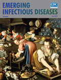
A Tale of Two Kitchens, Meals and Microbes [PDF - 2.76 MB - 2 pages]
| EID | Breedlove B, Meltzer MI. A Tale of Two Kitchens, Meals and Microbes. Emerg Infect Dis. 2018;24(6):1165-1166. https://doi.org/10.3201/eid2406.ac2406 |
|---|---|
| AMA | Breedlove B, Meltzer MI. A Tale of Two Kitchens, Meals and Microbes. Emerging Infectious Diseases. 2018;24(6):1165-1166. doi:10.3201/eid2406.ac2406. |
| APA | Breedlove, B., & Meltzer, M. I. (2018). A Tale of Two Kitchens, Meals and Microbes. Emerging Infectious Diseases, 24(6), 1165-1166. https://doi.org/10.3201/eid2406.ac2406. |


